
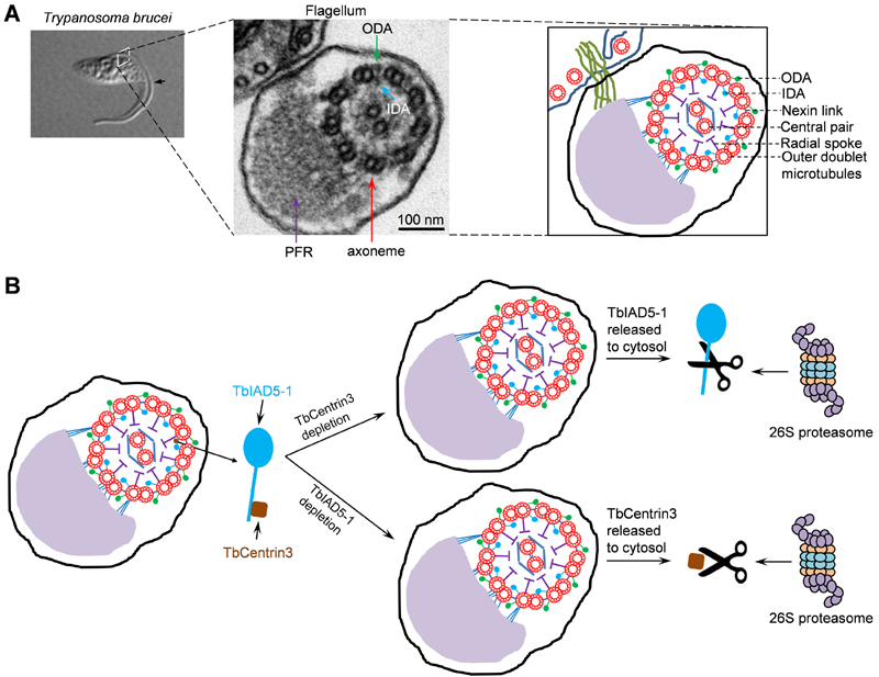
FIGURE 1: Role of TbCentrin3 in flagellar motility in trypanosomes.
(A) The trypanosome flagellum and its axoneme structure. Shown on the left panel is a differential interference contrast (DIC) image of a procyclic cell of T. brucei with a single flagellum (arrow). The middle panel shows the transmission electron microscopy of a cross-section of the flagellum and the cell body to which the flagellum is attached. The paraflagellar rod (PFR), the axoneme, outer dynein arm (ODA), and inner dynein arm (IDA) are indicated. The right panel is a schematic representation of the micrograph shown in the middle panel.
(B) Effect of TbCentrin3 depletion on the assembly and stability of TbIAD5-1 and vice versa. Note that only one IDA is labeled as the TbIAD5-1-TbCentrin3 complex and that TbCentrin3 and TbIAD5-1 are not drawn to the same scale. The precise numbers of inner dynein arms containing TbCentrin3 and TbIAD5-1 are still unclear. In the absence of TbCentrin3, newly synthesized TbIAD5-1 is not assembled to the axoneme and the old, axoneme-associated TbIAD5-1 is released to the cytosol, where it is degraded by the 26S proteasome. Conversely, in the absence of TbIAD5-1, new TbCentrin3 also fails to assemble to the axoneme and old TbCentrin3 is released from the axoneme to the cytosol, where it is degraded by the 26S proteasome.