
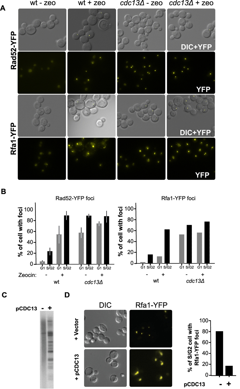
FIGURE 2: Rad52p and Rfa1p proteins form DNA damage foci in untreated cdc13Δ cells.
(A) Wild-type (wt) or cdc13Δ cells expressing the Rfa1-YFP fusion protein (RWY801) or the Rad52-YFP protein (CPY821) were analysed by fluorescence microscopy untreated or treated with zeocin for 3 h. Representative examples of the images obtained are shown.
(B) Quantification of cells with foci. Cells were morphologically divided into G1 cells (unbudded cells) and G2M cells (large budded cells). The Rad52-YFP experiment was performed in 3 independent biological replicates, and the Rfa1-YFP experiment was performed with at least 150 cells for each condition.
(C) Southern blot with DNA derived from MLY122 using a telomeric DNA probe. Cells were growing in the absence of Cdc13p (lane marked -) or the wt CDC13 gene was introduced via plasmid pCDC13 (lane marked +). Note that re-introduction of the protein CDC13 restores survivor type II TRF phenotype.
(D) The re-expression of CDC13 eliminates the formation of Rfa1-YFP foci in cdc13Δ cells. Strain CPY821 was grown to become Cdc13-independent and then an empty vector or pCDC13 was introduced. Representative examples of images obtained as in (A) are shown in the left panel. Quantification of the data is shown in the right panel. >100 cells were counted for each sample.