
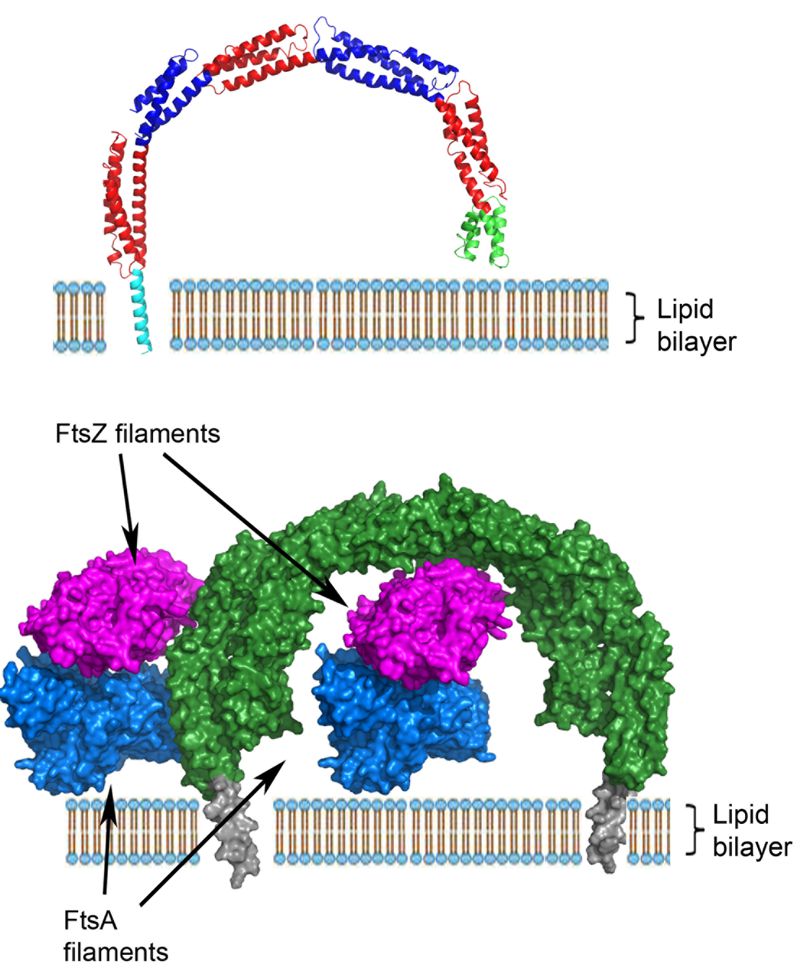
FIGURE 2:
Top – Model of the integration of B. subtilis EzrA into the lipid bilayer. The modelled N-terminal helix is coloured cyan, the remainder of the cytoplasmic domain is coloured as in Figure 1.
Bottom – Model for the interaction of EzrA (green, with the modelled TM helix grey) with FtsZ protofilaments (magenta) and the FtsZ membrane anchor, FtsA (blue) within the divisome; the FtsZ and FtsA filaments are viewed along the long axis of the respective filaments. EzrA is represented in an anti-parallel dimer that is observed in the crystal lattice and is supported by in vivo two hybrid studies. There is sufficient space to accommodate both FtsA and FtsZ in the gap between the membrane and the inside of the arch-shaped EzrA molecule.