
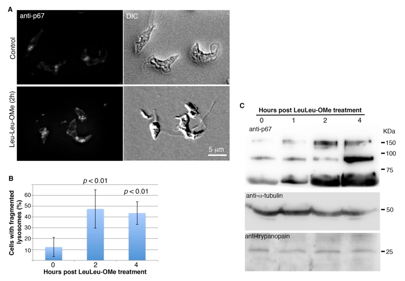
FIGURE 2: Lysosome destabilizing effects of LeuLeu-OMe.
(A) T. brucei cells were incubated with or without 30 µM LeuLeu-OMe, and samples were taken at t = 0 h, 1 h, 2 h and 4 h for immunofluorescence assays with anti-p67. Representative images of control and LeuLeu-OMe-treated cells are maximum intensity projection of serial optical sections through the entire cells.
(B) Quantitation of images as shown in (A) revealed an increase in lysosome fragmentation at 2 h and 4 h post Leuleu-OMe treatment. The quantitation results are presented as mean ± SD from 3 independent experiments.
(C) Samples treated as described in (A) were also processed for immunoblotting analyses with anti-p67 and anti-trypanopain. Anti-α-tubulin was used as a loading control.