
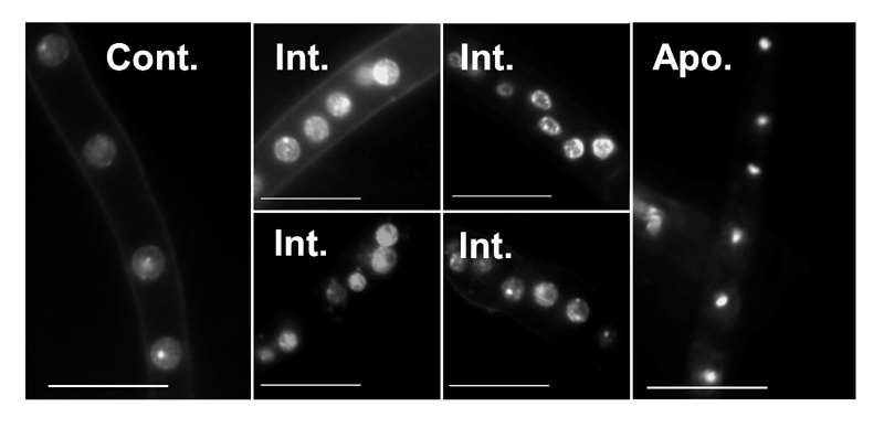
FIGURE 3: Changes in nuclei morphology in response to PCD-inducing conditions.
Cultures were grown for 24 h in PDB, then H2O2 was added to a final concentration of 10 mM. The mycelia were harvested after 4 h of incubation with H2O2, processed and stained with Hoechst 33342. Samples were visualized at 1000x magnification.
Control, time zero, cells are loosely and evenly stained, the nucleolus is clearly visible (arrow); Int., intermediate stages of apoptosis, accumulation of nuclear fragments is visible; Apoptotic, highly condensed nuclei represent the final stage of apoptosis. Scale bar = 10 µm.