
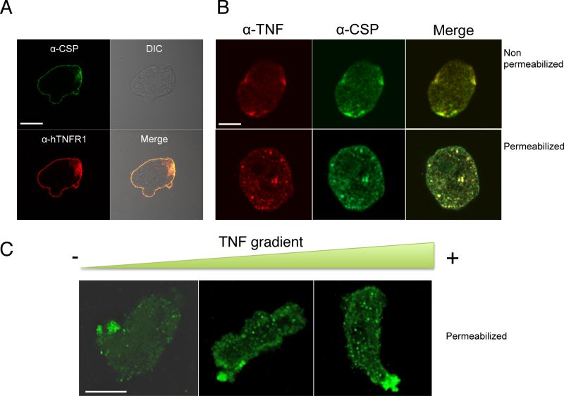
FIGURE 3: Cellular localization of CSP in Entamoeba histolytica.
(A) Immunolocalization of CSP (green) and hTNFR1 (red) in fixed but non-permeabilized trophozoites revealed that both proteins are located at the plasma membrane. Scale bar: 10 µm.
(B) Immunolocalization of CSP in E. histolytica incubated with a homogenous (non-gradient) concentration of TNF. Trophozoites were incubated with 70 nM TNF, fixed and permeabilized or not. CSP (green) and TNF (red) were co-localized at the trophozoite surface. CSP was detected as described for Fig. 3A. TNF was detected with 1E12 monoclonal antibody and mouse-Cy3 (dilution: 1:200). Scale bar: 10 µm.
(C) Immunolocalization of CSP in E. histolytica incubated with a TNF gradient. Trophozoites exposed to a TNF gradient for 2 h were fixed and permeabilized. During trophozoite migration toward the TNF source, CSP (green) concentrated in the rear part of the cells (i.e. distal to the TNF source). The nucleus was stained with DAPI (blue). Scale bar: 10 µm.