
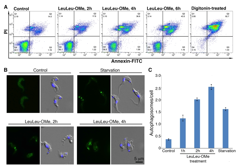
FIGURE 5: LeuLeu-OMe induces necrotic cell death and autophagy.
(A) Cells treated with 30 µM LeuLeu-OMe were fixed and stained with PI and annexin-FITC, and analyzed by flow cytometry. Digitonin-permeabilized cells were used as a positive control for the stains.
(B, C) Cell stably expressing YFP-TbATG8.2 were cultivated in medium with or without 30 µM LeuLeu-OMe. Autophagosome formation was monitored by relocalization of YFP-TbATG8.2 from a cytoplasmic distribution in control cells to a punctate structures using fluorescence microscopy. Autophagosome formation was quantitated and the results are shown as mean ± SD from 3 independent experiments. Cells starved in cytomix for 2 h were used as a positive control.