
Back to article: Evolution of the bacterial nucleosidase PpnN and its relation to the stringent response
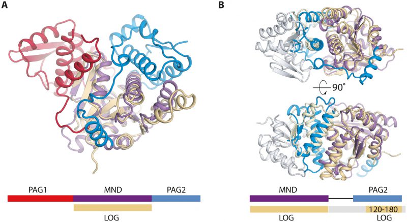
FIGURE 2: Structual comparison of E. coli PpnN and a LOG protein (A. thaliana lysine decarboxylase-like protein, PDB ID 2A33). (A) Comparison between a monomer of PpnN (purple, red, and blue) and LOG (beige) shows that the Rossmann-like fold is shared between the two families. Schematic in same colors below the structure for emphasis. (B) Superposition of a LOG dimer (grey and beige) onto the MND and PAG2 domain of PpnN reveals conserved secondary elements between LOG and PAG2 (yellow), more precisely two α-helices and two terminal β-strands. Schematic in same colour below show where the LOG dimer share secondary structure (beige) and the rest is coloured in grey.