
Back to article: Endomembrane remodeling and dynamics in Salmonella infection
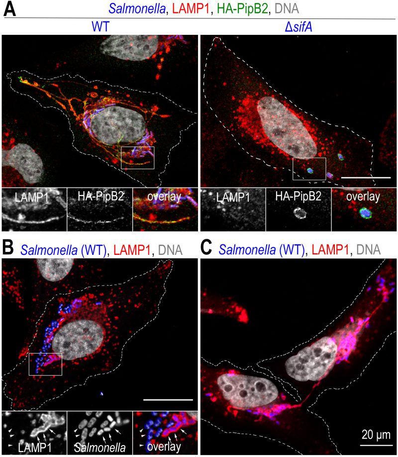
FIGURE 2: Salmonella induces a reorganization of the lysosomal compartment of the host cell. Confocal microscopy images, after immunolabelling, illustrating phenotypes observed in HeLa cells at a late time (12 h) of infection. (A) In cells infected with wild-type Salmonella (WT), the presence of bacteria (blue) in the juxtanuclear region is observed. The bacteria are in a vacuolar compartment (SCV) with a membrane similar to that of lysosomes, illustrated here by LAMP1 labelling (red). Membrane tubules (SITs) stretching to the cell periphery are very characteristic of Salmonella-infected HeLa cells. Some effectors secreted by T3SS-2, here HA-tagged PipB2 (green), are present on SCVs and SITs. In the absence of SifA (ΔsifA), the lysosome distribution of the infected cell appears to be little or not at all impaired. SCVs, which very often enclose several ΔsifA bacteria, adopt a position midway between the nucleus and the plasma membrane and are characterized by very weak LAMP1 labelling but strong labelling for membrane effectors (PipB2). (B) Salmonella is commonly found in the cytosol of epithelial cells. The image shows bacteria that are not surrounded by LAMP1 labelling (arrowheads) and are larger than those present in SCVs (arrows). (C) The image illustrates the distribution of SCVs in the two sister cells following mitosis and the fact that a SIT connects the two groups of bacteria until the two cells separate. Scale bar, 20 or 10 μm for the magnified insets. This figure is composed of unpublished images.