
Back to article: Endomembrane remodeling and dynamics in Salmonella infection
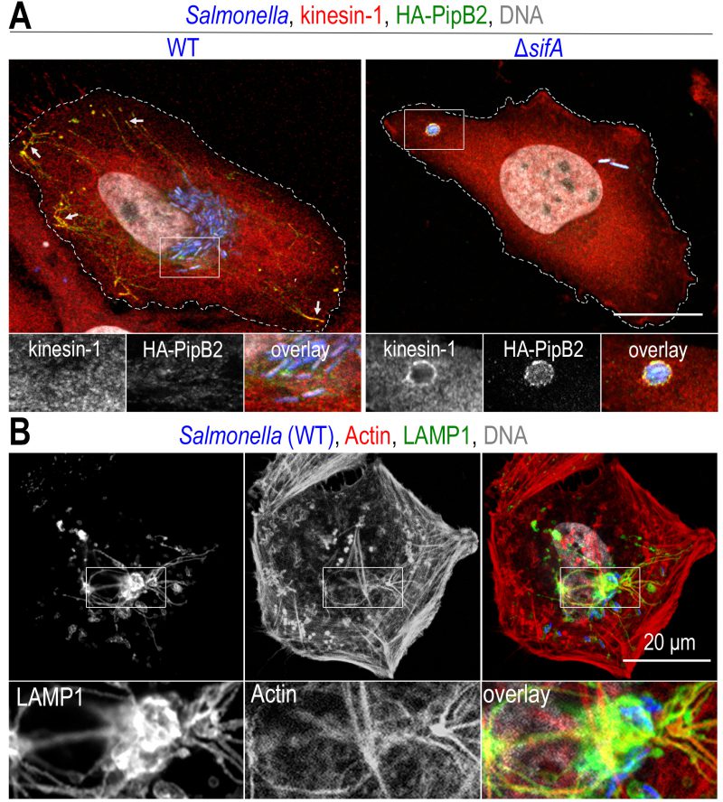
FIGURE 4: Recruitment of molecular motors and the actin cytoskeleton on Salmonella vacuoles. Confocal microscopy images, after immunolabelling, of Salmonella-infected HeLa (A) or Cos-7 (B) cells at a late time (12h) of infection. (A) Two effectors of T3SS-2, SifA and PipB2 recruit kinesin-1 to SCVs and SITs. The presence of kinesin-1 (red) and PipB2 (green) is detected at the tip of SITs (arrows) in cells infected with wild-type (WT) Salmonella (blue), while these molecules are difficult to detect on SCVs (see inset). In the absence of SifA (ΔsifA), kinesin-1 and membrane effectors accumulate on SCVs. (B) Actin labelling (red) highlights the presence of a strong actin cortex. Actin is also present around the juxtanuclear Salmonella microcolony (blue) and also marks the SIT network (LAMP1, in green). Scale bars, 20 or 10 μm for the magnified insets in (A) and 6.7 μm for the magnified insets in (B). This figure is composed of unpublished images.