
Back to article: A chemical genetic screen reveals a role for proteostasis in capsule and biofilm formation by Cryptococcus neoformans
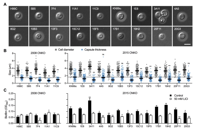
FIGURE 6: The ubiquitin/proteasome system contributes to capsule and biofilm formation. (A) DIC microscopy images of the indicated ubiquitin/proteasome-associated C. neoformans strains (see Table 1) grown in CIM for 48 h and stained with India ink to visualize capsule via dye exclusion. H99C, Wt control for strains from the 2008 CNKO collection. KN99a, Wt control for strains from the 2015 CNKO collection. Scale bar, 5 µm. (B) Quantification of cell diameter (grey squares) and capsule thickness (blue circles) for cells from panel A. The experiment was performed twice, and at least 100 cells were analyzed per strain and condition. The black bar indicates the median ± interquartile range. The dotted black lines indicate the median capsule sizes of the respective Wt controls. ns, not significant. **P < 0.01 by two-way ANOVA compared to the respective Wt. (C) Biofilm formation by ubiquitin/proteasome-associated mutants. Indicated strains were grown under biofilm-inducing conditions without (control) or with 50 mM LiCl and quantified by the XTT reduction assay. OD492, optical density at 492 nm. Results are the mean ± SEM of three independent experiments, each performed in triplicate. * P < 0.05, and **P < 0.01 by t-test compared to the respective Wt growth condition.