
Back to article: A comparative approach to decipher intestinal animal-microbe associations
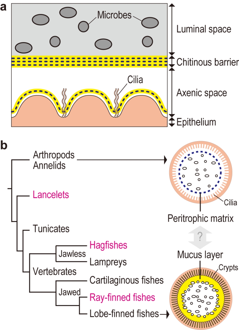
FIGURE 1: Chitin-based barrier immunity. (a) A model for the tunicate Ciona intestinalis Type A. The gut space is radially compartmentalized into luminal space, to which filter-fed microbes are confined, and peripheral axenic space over the ciliated epithelium, defined by multi-layered chitinous membranes. Chitin-ous membranes are framed with meshed chitin nanofibers (blue dotted line) embedded in proteinaceous matrix containing gel-forming mucin (yellow). Delamination of a new membrane from the epithelium renews axenic conditions. (b) Evolutionary im-plications. The tree diagram at left depicts phylogenetic relationships of chordates. Two arrows extending from invertebrate outgroups and lobe-finned fishes, which include mammals, point to schematic drawings of intestinal barrier immunity representative of each group. The Ciona chitinous barrier (a) provides molecular evidence for a possible link between the invertebrate peritrophic matrix (blue dotted line) and the mammalian mucus layer (yellow). To test this idea, animal groups that occupy intervening phylogenetic positions (typed in magenta) are critical. Adapted from Nakashima et al. Nat Commun 9: 3402.