
Back to article: A simple microfluidic platform to study age-dependent protein abundance and localization changes in
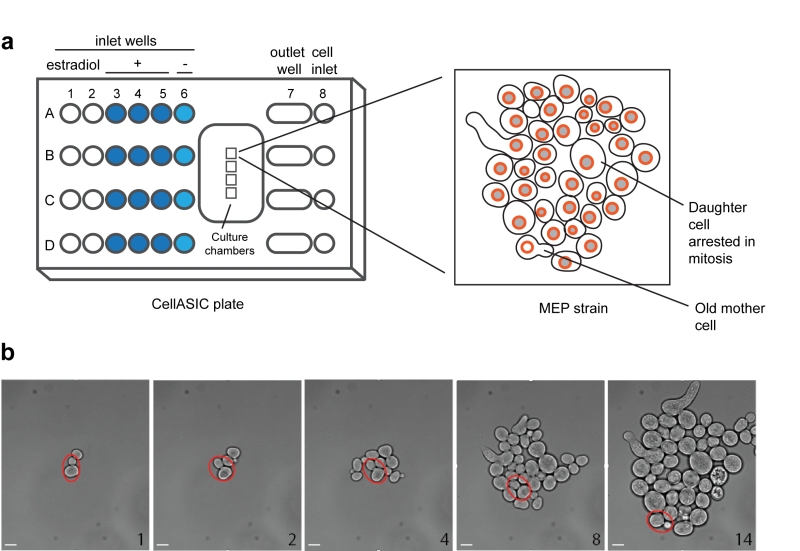
Figure 1: Design of a CellASIC-based platform to study replicative yeast ag-ing. (a) Four strains (A-D) can be monitored for long periods using a CellASIC microfluidic system. Once cells are trapped in the culture chambers, perfusion of the media takes place for 60 min and perfusion of the media with estradiol is active for 34 h. After addition of estradiol, only the daughter cells of the MEP strain will be arrested in mitosis and cannot proliferate whereas the mother cell continues dividing. Input wells 1 and 2 are not needed for these experiments and are left empty. (b) Bright-field images show the growth of a colony from a founder mother cell (circled in red). Scale bar, 5 µm.