
Back to article: Chlamydia and mitochondria - an unfragmented relationship
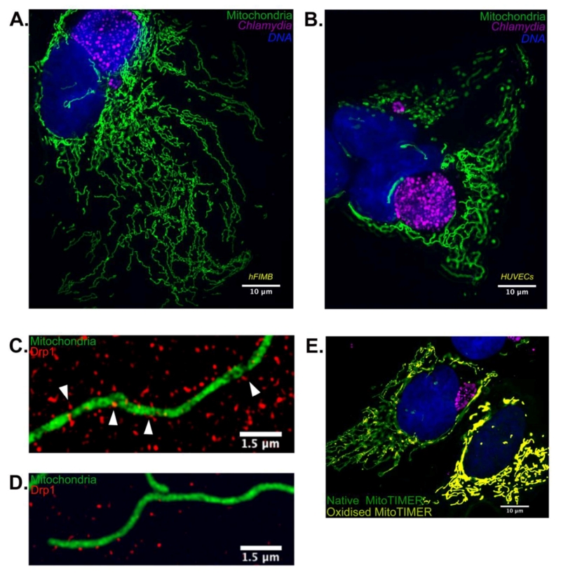
FIGURE 1: Chlamydia infection alters mitochondrial morphology in primary cells by inhibition of Drp1 expression. (A) 36 hours of Chlamydia infec-tion in hFIMB cells lead to an abnormal increase in mi-tochondrial fragment length while preventing H2O2 mediated fragmenta-tion. The sample was treated with 1μM H2O2 for 1 hour. (B) In this panel both the HUVECs have been treated with CoCl2 for 6 hours, however, only the cell harboring a very early stage Chlamydia inclusion (8 hours) undergoes severe mitochondrial fragmentation. Advanced stages of Chlamydia infection (36 hours; right most cell) grants the host cell mitochondria protection against extrinsic pro-fragmentation stress mediated by CoCl2. Fragments of mitochondria from (C) non-infected and (D) Chlamydia-infected HUVECs decorated with Drp1 aggregates. The white arrows in (C) indicate successful Drp1 aggregate asso-ciation with the mitochondria and initiation of fission. (E) HUVECs transfected with Mito-TIMER and infected with Chlamydia for 20 hours. The non-infected cell (right most) exhibits a drastic increase in the red form (oxidized form) of the MitoTIMER protein along with a decrease in mito-chondrial fragment length.