
Back to article: Uncovering the hidden: complexity and strategies for diagnosing latent tuberculosis
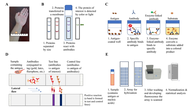
FIGURE 1: Schematic representation of how diagnostic methods have evolved to detect TB infection. (A) The Tuberculin Skin Test (also known as Mantoux test) depends on the inflammatory response, mediated by immune cells influx, localized at the site of injection in the forearm; (B) detection of antigenic proteins by a technique called “Western blot”, where antibodies are present in the blood of a suspected TB-infected person, and detect antigens present in a membrane; (C) another approach that detects antigens via antibodies is the “ELISA” test; this is used, for example in the Interferon gamma release assays. The difference between “Western blot” and “ELISA” assays is where the reaction takes places, in a matrix or in solution, respectively; (D) a variation of “Western blot” is the “Lateral flow assay”, where multiple antigen-antibody reaction occurs simultaneously in a membrane, and these are detected as colored bands; (E) finally, the whole set of protein or nucleic acids sequences can be screened via hybridization and using fluorescent labels. Considering the vast number of reactions occurring here, normalization and statistical analyses are conducted to ascertain what signal is real and not background/noise and therefore not relevant.