Back to article: Promoter methylation and increased expression of PD-L1 in patients with active tuberculosis
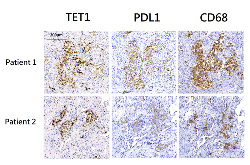
FIGURE 3: Immunohistochemistry (IHC) staining of lung tissues from two patients with pulmonary tuberculosis. IHC staining using anti-PD-L1 and anti-TET1 antibodies showed the presence of PD-L1 and TET1 positive cells. It also demonstrated the co-localization of PD-L1 and TET1. IHC staining using an anti-CD68 antibody suggested that these PD-L1/TET1 positive cells were primarily macrophages. PD-L1 – programmed death-ligand 1; TET1 – ten eleven translocation methylcytosine dioxygenases.

