
Back to article: Pathogenic Escherichia coli change the adhesion between neutrophils and endotheliocytes in the experimental bacteremia model
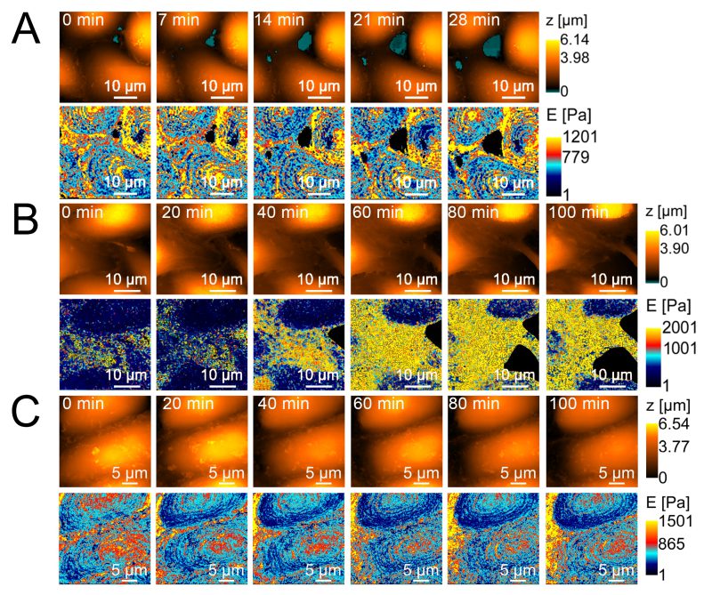
FIGURE 4: Morphology and stiffness of endothelial cells assessed by scanning ion-conductance microscopy. (A) control; top line – morphology, bottom line – stiffness (dynamics taken at 7 min intervals); (B) after application of pathogenic strain E. coli 321; top line – morphology, bottom line – stiffness (dynamics taken at 20 min intervals); (C) – after application of non-pathogenic strain E. coli HB101; top line – morphology, bottom line – stiffness (dynamics taken at 20 min intervals).