Back to article: Microwave-assisted preparation of yeast cells for ultrastructural analysis by electron microscopy
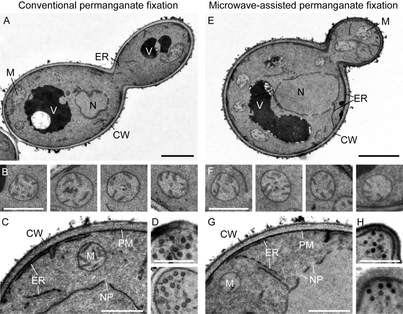
FIGURE 1: TEM of Saccharomyces cerevisiae fixed with potassium permanganate. (A) Wild type yeast cells were grown to logarithmic growth phase in galactose-containing rich medium and prepared for electron microscopy by conventional permanganate fixation (protocols 1 and 3). (B, C) Mitochondria, cell cortex and nuclear envelope in cross sections of wild type yeast cells prepared by conventional permanganate fixation. (D) Secretory vesicles at small emerging buds of cells prepared as above. (E) Wild type yeast cells were grown as in (A) and prepared for electron microscopy by microwave-assisted permanganate fixation (protocols 2 and 3). (F-H) Organelles in cross sections of wild type yeast cells prepared by microwave-assisted permanganate fixation. CW, cell wall; ER, endoplasmic reticulum; M, mitochondrion; N, nucleus; NP, nuclear pore; PM, plasma membrane; V, vacuole. Black bars, 1 µm; white bars, 500 nm.

