Table of contents

Volume 6, Issue 2, pp. 105 - 141, February 2019

Issue cover
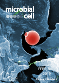
Cover: Digitally colorized scanning electron microscopic (SEM) image of an untreated water specimen extracted from a wild stream mainly used to control flooding during inclement weather, revealed the presence of unidentified organisms, which included bacteria, protozoa, and algae. In the center of this image was an exquisitely formed, unidentified, round vesicle-shaped microorganism, which may have been algal, or diatomic. Image by Janice Haney Carr (Centers for Disease Control and Prevention, USA; Public Health Image Library, image ID #11708); image modified by MIC. The cover is published under the Creative Commons Attribution (CC BY) license. Enlarge issue cover

Reviews

The extracellular matrix of mycobacterial biofilms: could we shorten the treatment of mycobacterial infections?

Poushali Chakraborty and Ashwani Kumar

page 105-122 | 10.15698/mic2019.02.667 | Full text | PDF | Abstract

A number of non-tuberculous mycobacterium species are opportunistic pathogens and ubiquitously form biofilms. These infections are often recalcitrant to treatment and require therapy with multiple drugs for long duration. The biofilm resident bacteria also display phenotypic drug tolerance and thus it has been hypothesized that the drug unresponsiveness in vivo could be due to formation of biofilms inside the host. We have discussed the biofilms of several pathogenic non-tuberculous mycobacterium (NTM) species in context to the in vivo pathologies. Besides pathogenic NTMs, Mycobacterium smegmatis is often used as a model organism for understanding mycobacterial physiology and has been studied extensively for understanding the mycobacterial biofilms. A number of components of the mycobacterial cell wall such as glycopeptidolipids, short chain mycolic acids, monomeromycolyl diacylglycerol, etc. have been shown to play an important role in formation of pellicle biofilms. It shall be noted that these components impart a hydrophobic character to the mycobacterial cell surface that facilitates cell to cell interaction. However, these components are not necessarily the constituents of the extracellular matrix of mycobacterial biofilms. In the end, we have described the biofilms of Mycobacterium tuberculosis (Mtb), the causative agent of tuberculosis. Three models of Mtb biofilm formation have been proposed to study the factors regulating biofilm formation, the physiology of the resident bacteria, and the nature of the biomaterial that holds these bacterial masses together. These models include pellicle biofilms formed at the liquid-air interface of cultures, leukocyte lysate-induced biofilms, and thiol reductive stress-induced biofilms. All the three models offer their own advantages in the study of Mtb biofilms. Interestingly, lipids (mainly keto-mycolic acids) are proposed to be the primary component of extracellular polymeric substance (EPS) in the pellicle biofilm, whereas the leukocyte lysate-induced and thiol reductive stress-induced biofilms possess polysaccharides as the primary component of EPS. Both models also contain extracellular DNA in the EPS. Interestingly, thiol reductive stress-induced Mtb biofilms are held together by cellulose and yet unidentified structural proteins. We believe that a better understanding of the EPS of Mtb biofilms and the physiology of the resident bacteria will facilitate the development of shorter regimen for TB treatment.

In the pit

What’s in a name? How organelles of endosymbiotic origin can be distinguished from endosymbionts

Ansgar Gruber

page 123-133 | 10.15698/mic2019.02.668 | Full text | PDF | Abstract

Mitochondria and plastids evolved from free-living bacteria, but are now considered integral parts of the eukaryotic species in which they live. Therefore, they are implicitly called by the same eukaryotic species name. Historically, mitochondria and plastids were known as “organelles”, even before their bacterial origin became fully established. However, since organelle evolution by endosymbiosis has become an established theory in biology, more and more endosymbiotic systems have been discovered that show various levels of host/symbiont integration. In this context, the distinction between “host/symbiont” and “eukaryote/organelle” systems is currently unclear. The criteria that are commonly considered are genetic integration (via gene transfer from the endosymbiont to the nucleus), cellular integration (synchronization of the cell cycles), and metabolic integration (the mutual dependency of the metabolisms). Here, I suggest that these criteria should be evaluated according to the resulting coupling of genetic recombination between individuals and congruence of effective population sizes, which determines if independent speciation is possible for either of the partners. I would like to call this aspect of integration “sexual symbiont integration”. If the partners lose their independence in speciation, I think that they should be considered one species. The partner who maintains its genetic recombination mechanisms and life cycle should then be the name giving “host”; the other one would be the organelle. Distinguishing between organelles and symbionts according to their sexual symbiont integration is independent of any particular mechanism or structural property of the endosymbiont/host system under investigation.

In the beginning was the word: How terminology drives our understanding of endosymbiotic organelles

Miroslav Oborník

page 134-141 | 10.15698/mic2019.02.669 | Full text | PDF | Abstract

The names we give objects of research, to some extent, predispose our ways of thinking about them. Misclassifications of Oomycota, Microsporidia, Myxosporidia, and Helicosporidia have obviously affected not only their formal taxonomic names, but also the methods and approaches with which they have been investigated. Therefore, it is important to name biological entities with accurate terms in order to avoid discrepancies in researching them. The endosymbiotic origin of mitochondria and plastids is now the most accepted scenario for their evolution. Since it is apparent that there is no natural definitive border between bacteria and semiautonomous organelles, I propose that mitochondria and plastids should be called bacteria and classified accordingly, in the bacterial classification system. I discuss some consequences of this approach, including: i) the resulting “changes” in the abundances of bacteria, ii) the definitions of terms like microbiome or multicellularity, and iii) the concept of endosymbiotic domestication.

By continuing to use the site, you agree to the use of cookies. more information

The cookie settings on this website are set to "allow cookies" to give you the best browsing experience possible. If you continue to use this website without changing your cookie settings or you click "Accept" below then you are consenting to this. Please refer to our "privacy statement" and our "terms of use" for further information.

Close