
Back to issue: March 2017
Microbial Cell – March 2017
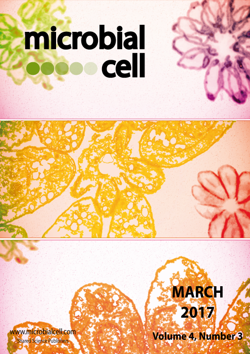
Combination of (i) fluorescense images of intracellular Toxoplasma gondii tachyzoites showing division process by endodyogeny. Parasites were stained with anti-GAP45 for mother cell pellicle, anti-IMC1 for daughter and mother cell pellicle and DAPI for nucleus and (ii) an electron micrograph of an intracellular rosette of Toxoplasma gondii (image by Erica dos Santos Martins-Duarte and Wanderley de Souza, Universidade Federal do Rio de Janeiro, Brazil); image modified by MIC. The cover is published under the Creative Commons Attribution (CC BY) license.