Table of contents
Volume 6, Issue 9, pp. 373 - 453, September 2019
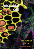
Cover: The image shows a mouse colon section of the lumen, showing fiber elements (yellow and orange), the mucus (green, UEA-1 stain) and bacteria (magenta and green). Image by Carolina Tropini, University of British Columbia (Canada) and Justin Sonnenburg, Stanford University (USA); image modified by MIC. The cover is published under the Creative Commons Attribution (CC BY) license.
Enlarge issue cover
Beyond cells – The virome in the human holobiont
Rodrigo García-López, Vicente Pérez-Brocal and Andrés Moya
Reviews |
page 373-396 | 10.15698/mic2019.09.689 | Full text | PDF |
Abstract
Viromics, or viral metagenomics, is a relatively new and burgeoning field of research that studies the complete collection of viruses forming part of the microbiota in any given niche. It has strong foundations rooted in over a century of discoveries in the field of virology and recent advances in molecular biology and sequencing technologies. Historically, most studies have deconstructed the concept of viruses into a simplified perception of viral agents as mere pathogens, which demerits the scope of large-scale viromic analyses. Viruses are, in fact, much more than regular parasites. They are by far the most dynamic and abundant entity and the greatest killers on the planet, as well as the most effective geo-transforming genetic engineers and resource recyclers, acting on all life strata in any habitat. Yet, most of this uncanny viral world remains vastly unexplored to date, greatly hindered by the bewildering complexity inherent to such studies and the methodological and conceptual limitations. Viromic studies are just starting to address some of these issues but they still lag behind microbial metagenomics. In recent years, however, higher-throughput analysis and resequencing have rekindled interest in a field that is just starting to show its true potential. In this review, we take a look at the scientific and technological developments that led to the advent of viral and bacterial metagenomics with a particular, but not exclusive, focus on human viromics from an ecological perspective. We also address some of the most relevant challenges that current viral studies face and ponder on the future directions of the field.
Diverse conditions support near-zero growth in yeast: Implications for the study of cell lifespan
Jordan Gulli, Emily Cook, Eugene Kroll, Adam Rosebrock, Amy Caudy and Frank Rosenzweig
Reviews |
page 397-413 | 10.15698/mic2019.09.690 | Full text | PDF |
Abstract
Baker’s yeast has a finite lifespan and ages in two ways: a mother cell can only divide so many times (its replicative lifespan), and a non-dividing cell can only live so long (its chronological lifespan). Wild and laboratory yeast strains exhibit natural variation for each type of lifespan, and the genetic basis for this variation has been generalized to other eukaryotes, including metazoans. To date, yeast chronological lifespan has chiefly been studied in relation to the rate and mode of functional decline among non-dividing cells in nutrient-depleted batch culture. However, this culture method does not accurately capture two major classes of long-lived metazoan cells: cells that are terminally differentiated and metabolically active for periods that approximate animal lifespan (e.g. cardiac myocytes), and cells that are pluripotent and metabolically quiescent (e.g. stem cells). Here, we consider alternative ways of cultivating Saccharomyces cerevisiae so that these different metabolic states can be explored in non-dividing cells: (i) yeast cultured as giant colonies on semi-solid agar, (ii) yeast cultured in retentostats and provided sufficient nutrients to meet minimal energy requirements, and (iii) yeast encapsulated in a semisolid matrix and fed ad libitum in bioreactors. We review the physiology of yeast cultured under each of these conditions, and explore their potential to provide unique insights into determinants of chronological lifespan in the cells of higher eukaryotes.
The multiple functions of the numerous Chlamydia trachomatis secreted proteins: the tip of the iceberg
Joana N. Bugalhão and Luís Jaime Mota
Reviews |
page 414-449 | 10.15698/mic2019.09.691 | Full text | PDF |
Abstract
Chlamydia trachomatis serovars are obligate intracellular bacterial pathogens mainly causing ocular and urogenital infections that affect millions of people worldwide and which can lead to blindness or sterility. They reside and multiply intracellularly within a membrane-bound vacuolar compartment, known as inclusion, and are characterized by a developmental cycle involving two morphologically and physiologically distinct chlamydial forms. Completion of the developmental cycle involves the secretion of > 70 C. trachomatis proteins that function in the host cell cytoplasm and nucleus, in the inclusion membrane and lumen, and in the extracellular milieu. These proteins can, for example, interfere with the host cell cytoskeleton, vesicular and non-vesicular transport, metabolism, and immune signalling. Generally, this promotes C. trachomatis invasion into, and escape from, host cells, the acquisition of nutrients by the chlamydiae, and evasion of cell-autonomous, humoral and cellular innate immunity. Here, we present an in-depth review on the current knowledge and outstanding questions about these C. trachomatis secreted proteins.
Evolution of the bacterial nucleosidase PpnN and its relation to the stringent response
René Lysdal Bærentsen, Ditlev Egeskov Brodersen and Yong Everett Zhang
Microreviews |
page 450-453 | 10.15698/mic2019.09.692 | Full text | PDF |
Abstract
In our recent publication (Zhang et al., 2019), we demonstrate an interesting mode of regulation of purine metabolism unique to Proteobacteria. In this microreview, we would like to reflect on the ideas put forward, with special focus on protein domain architecture of the enzyme involved, its orthologues in plants, and the implications of the differential effects observed between binding of the two alarmone molecules, ppGpp (guanosine 3′,5′-bisdiphosphate) and pppGpp (guanosine-5′-triphosphate-3′-diphosphate). In our previous work, we showed that the Escherichia coli nucleotide 5′-monophosphate nucleosidase, PpnN, which is conserved in Proteobacteria, cleaves its preferred substrate, guanosine monophosphate (GMP), at a much higher rate in the presence of both pppGpp and ppGpp (Figure 1A). Structural analysis reveals that binding of pppGpp leads to a conformational change in the protein that exposes its active site, suggesting this is the reason for the observed increase in activity. Finally, point mutation of the alarmone-interacting residues show a defect in binding, resulting in (i) increased basal catalytic activity of PpnN and higher competitive fitness of E. coli in an environment with fluctuating nutrient levels, and (ii) increased bacterial sensitivity towards antibiotics. In contrast, complete loss of the ppnN gene has the inverse effect, i.e. reduced competitive growth and improved antibiotic tolerance. We used these observations to propose a model in which E. coli uses PpnN to balance the need of fitness (fast growth) against tolerance towards antibiotics to improve survival.


