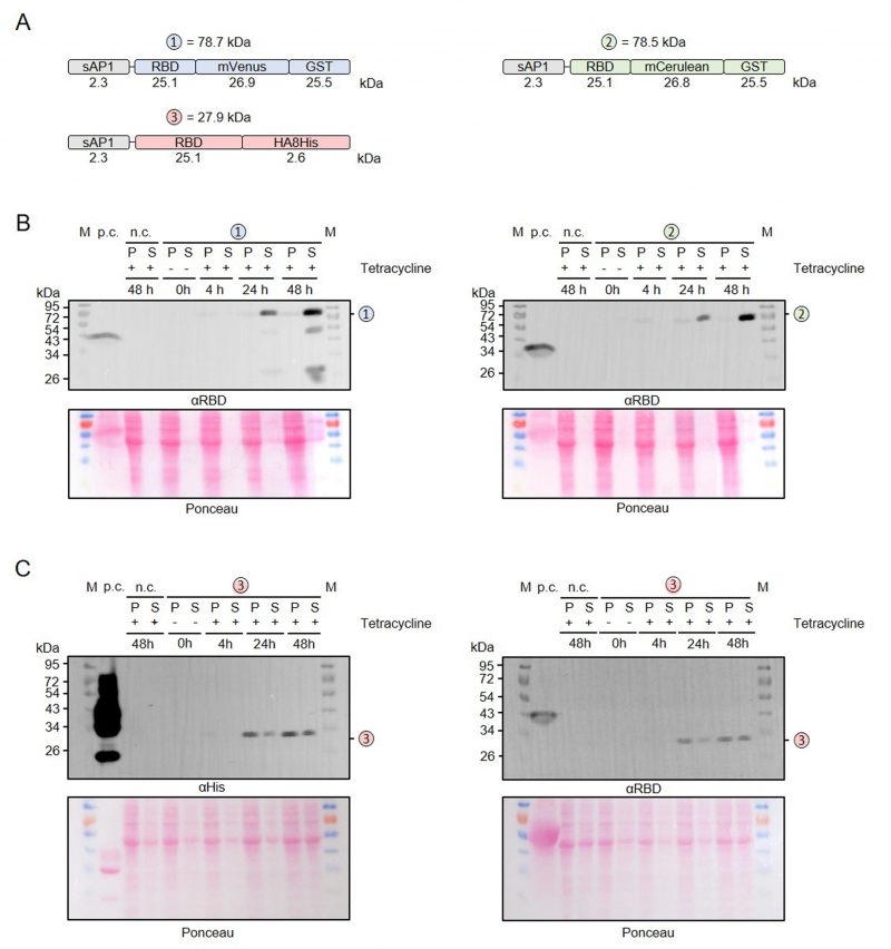
FIGURE 4: Time course measurement for the production and secretion of recombinant RBD. (A) Schematic overview and expected mass of the secreted RBD fusion proteins. (B) Western blot analysis with an antibody against the RBD domain of RBD fusion variants 1 and 2 in the cell-containing pellet fraction (P) and supernatant fraction (S) following tetracycline induction of the according L. tarentolae liquid cultures for the indicated time periods. An induced culture without plasmid served as negative control (n.c.) and recombinant RBD as positive control (p.c.). The calculated masses from panel A are indicated. (C) Western blot analysis with antibodies against the C-terminal His tag (left) or the RBD domain (right) of fusion variant 3.
By continuing to use the site, you agree to the use of cookies. more information
The cookie settings on this website are set to "allow cookies" to give you the best browsing experience possible. If you continue to use this website without changing your cookie settings or you click "Accept" below then you are consenting to this. Please refer to our "privacy statement" and our "terms of use" for further information.

