
Back to article: Chlamydia trachomatis’ struggle to keep its host alive
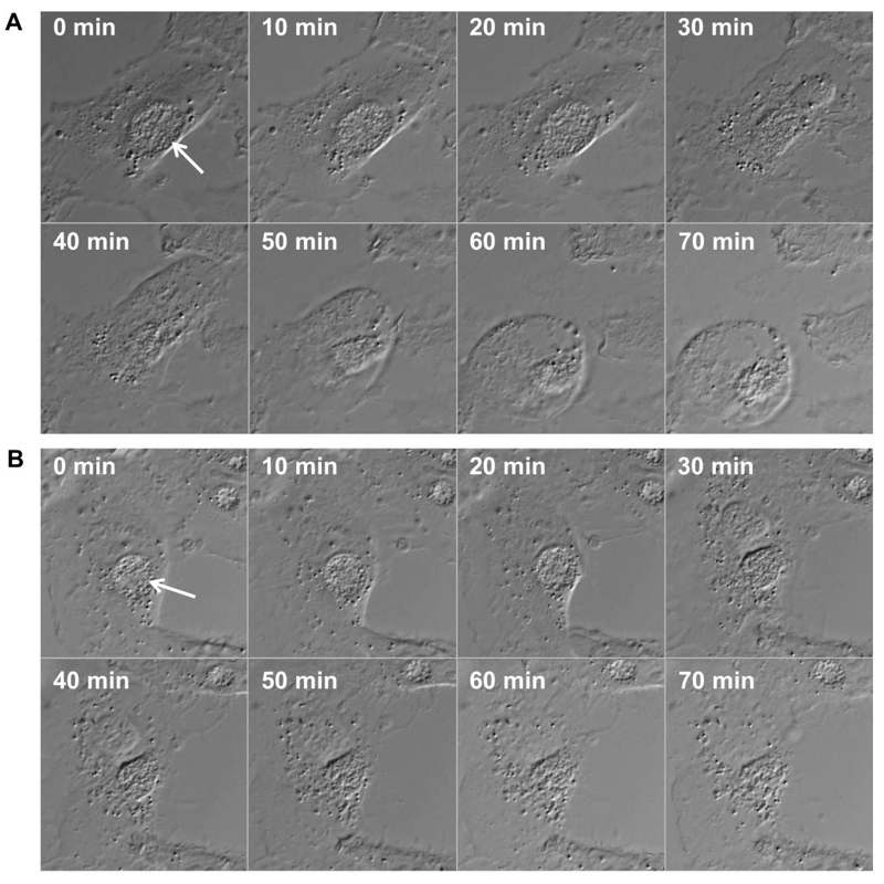
FIGURE 2: Time lapse microscopic image series displaying inclusion and plasma membrane rupture in HeLa cells infected with CpoS-deficient C. trachomatis.
HeLa cells (ATCC CCL-2) were infected with CpoS-deficient C. trachomatis L2/434/Bu (CTL2-cpoS::bla, 10 inclusion-forming units/cell) and imaged for over 40 hours in 10-min intervals at an Axio Observer.Z1 microscope (Zeiss). The displayed image series (A and B) document the process of necrotic cell death in two selected cells. The morphologic changes in these dying cells suggest that the rupture of the inclusion membrane occurred simultaneously to, or immediately before, the rupture of the plasma membrane. Arrows indicate inclusions.