
Manuscript Preparation
This section will assist you in preparing your article files. The graph below is an overview, which includes all necessary information to prepare your submission. Further down, you will find an extended description. Do not hesitate to contact us if you have any question.
Submission overview and guidelines
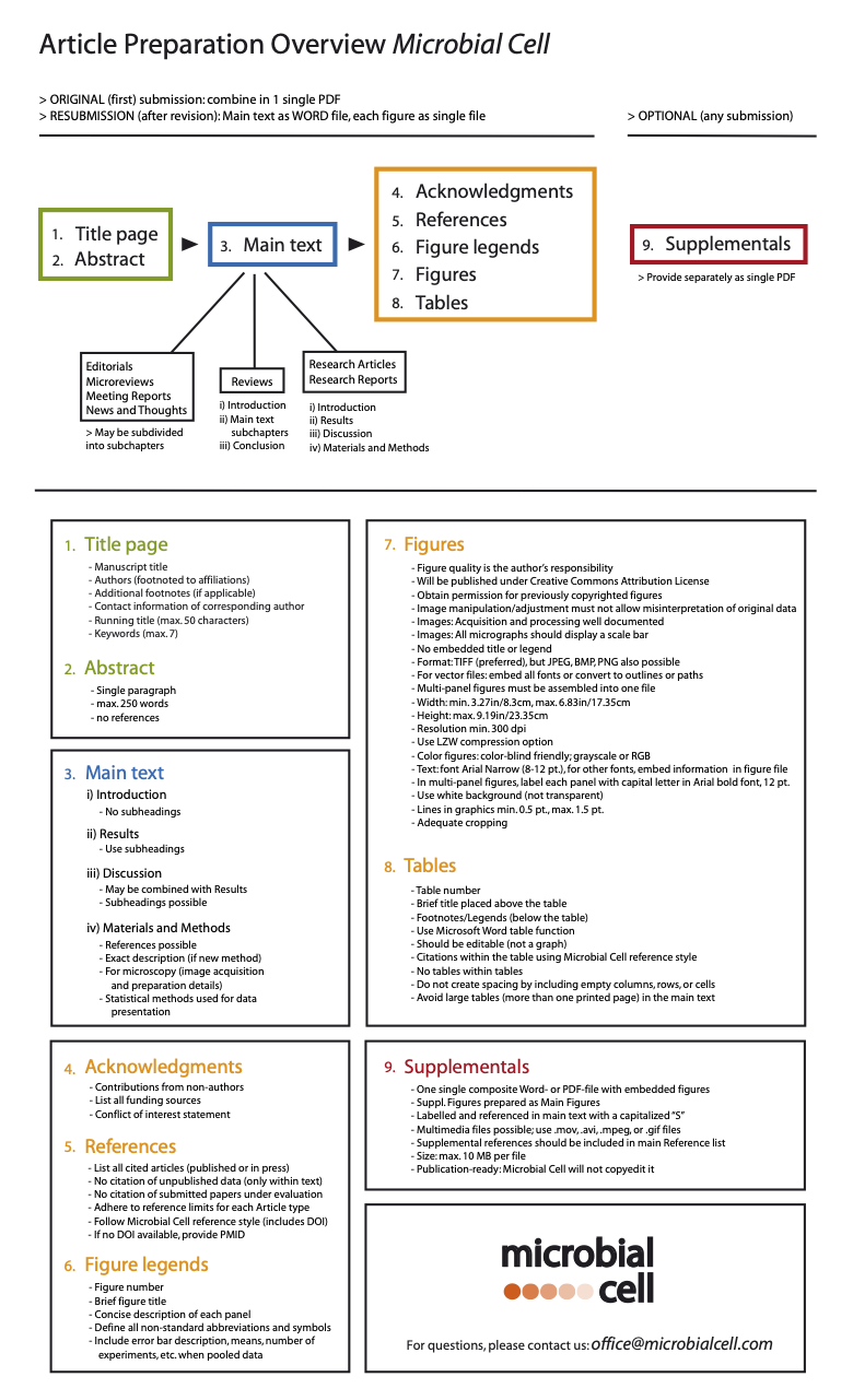
Please click on the graph below before preparing your manuscript: it includes all relevant information for the preparation of each subsection of your manuscript. You can also download the overview as PDF here.
If you have prepared your manuscript accordingly, please submit via our submission system:

Extended description
If you have any doubt regarding the preparation or submission of your manuscript, please read through the corresponding description in this section or contact us.
General guidelines
Preparation of specific manuscript sections
- General aspects
- Title page
- Abstract
- Main text
- Acknowledgments
- References
- Figure legends
- Microscopic images and gel images
- Figure preparation
- Tables
Special submission items
Submission
If it is an original submission, prepare a single PDF file including the main text (title through figure legends), all figures, and the supplemental data (in this order): this PDF file will be used for peer-review purposes and should not exceed 10 MB. In addition, provide the cover letter as a separate document (MS Word or PDF). If it is a resubmission (after revision), provide the cover letter, the point-by-point reply as well as the main text (title through figure legends) as single MS Word documents, respectively. In addition, provide each figure file as well as the supplemental material file separately.
Note that your files will be handled and organized at the editorial level upon receipt and that your cover letter will be seen by the handling editors only (i.e. not the referees).
Cover Letter
Upon submission, you should supply a cover letter (approximately one page) containing the following pieces of information:
– The corresponding author’s contact information.
– A concise summary of your study and a brief explanation of its impact.
– A specification of the article type you are submitting.
– You may suggest up to 3 suitable members of our Editorial Board for handling of your manuscript.
– A list of recommended reviewers (up to 5). Please supply their names, affiliations and e-mail addresses. If you would like to request the exclusion of potential reviewers from the review process, please provide their names (up to 3).
– In principle, Microbial Cell allows the deposition of a manuscript in a preprint server like BioRxiv; however, please mention this in the Cover Letter if applicable.
Language
We expect submitted manuscripts to be written in clear, comprehensible English. If you have concerns about your level of English, please have your manuscript proofread by a native English speaker or a professional scientific editing service prior to submission. This will ensure that reviewers are better able to read and assess your manuscript.
Word and Figure limits
Microbial Cell publishes a whole range of different Article Types, to which we apply very broad limits in terms of word count, number of figures or references. For these formal restrictions, please consult our article description and table overview under the Article Types section.
General Format Guidelines
The text should be double-spaced (i.e. double line spacing) and pages should be numbered. Appropriate standardized nomenclature should be employed, including the suitable use of species, gene and protein names as well as SI units. Abbreviations should be kept at a minimum. Non-standard abbreviations should be used only if appearing at least twice in the text and in that case be defined upon first usage. If you are using symbols or special characters, please do it by inserting them via the “Insert” tab (Insert → Symbols), but do NOT use the font “Symbol”. Thank you!
Submitted manuscripts that do not comply with the format guidelines will be returned to the authors for reformatting.
General aspects
All manuscripts independent of the article type should start with a Title Page and an Abstract ensued by the main text (in Microreviews the Abstract is the first paragraph of the main text). After the main text, the following sections should be included in this order if applicable: Acknowledgements, References, Figure and Table Legends, Tables. If it is an original submission, Figures and Supplementary Material should be integrated with the main text into a single PDF file to be used for peer-review purposes and should not exceed 10 MB. If it is a resubmission (after revision), each figure file as well as the supplemental material file should be provided separately.
The main text of “Editorials”, “Microreviews”, “Meeting Reports” as well as “News and Thoughts” manuscripts can be subdivided into different chapters depending on their length and the discussed topic at the discretion of the authors and editors. The main text of “Reviews” is divided into an (i) introduction, (ii) the main article body (that can be subdivided into different chapters) and (iii) a conclusion. The main text of “Research Articles” and “Research Reports” usually includes Introduction, Results, Discussion and Materials and Methods as separate sections. Please prepare the different sections in exactly this order. However, results and discussion may be combined in one single section if preferred.
Below, please find details on the preparation of the different sections:
Title page
The first page of your manuscript should include the following information:
– The TITLE, which should concisely describe the article’s conceptual significance and be accessible to the broad readership of Microbial Cell. It must be less than 150 characters (including spaces). – The AUTHORS, provided as a complete author name list (complete first and last names; middle name(s) may be provided as initial(s)). Each author should be footnoted to her/his corresponding affiliation(s). Please carefully double-check the correct spelling of the names, since changes in the post-production phase are laborious and may be implemented with substantial delay.
– The AFFILIATIONS, which should include the following information: department/subunit/institute; institution/university; city, state/province (if applicable), and country. Please carefully double-check the correct affiliation(s) for each author, since changes in the post-production phase are laborious and may be implemented with substantial delay.
– Any ADDITIONAL FOOTNOTES, which should be used if a present address or a statement of equal contribution (first and/or senior authorship) needs to be included.
– The CONTACT information, which should include the name, mailing address, telephone/fax numbers, and e-mail address of the corresponding author, i.e. the author with whom Microbial Cell’s editorial and production offices should maintain correspondence. If you would like to have two published corresponding authors, you still have to designate one of them for communication with Microbial Cell in all matters prior to publication. The published corresponding author(s) confirms adherence to all editorial and submission policies and is responsible for any communications that may result after publication.
– The RUNNING TITLE that must be 50 characters or less and should present the article’s topic.
– The KEYWORDS that should include up to 7 key general topics to which the article can be related.
For REVISED MANUSCRIPTS, please include the original manuscript number assigned upon original submission and the word “REVISION” in the title page.
Abstract
The Abstract should be a single paragraph of not more than 250 words that gives a clear synopsis of the article’s content and its significance. Shorter abstracts are encouraged. Abstracts should not include references.
Main text
Introduction
The Introduction should provide referenced background information and put the work’s purpose into context to allow the readership outside the specific field to understand why the study was performed. This section may include several paragraphs but no subheadings. You may want to conclude this section with a brief description of the work’s overall result and comment on its significance.
Results
The Results section should include all relevant data. It should be logically and constructively presented and be divided with subheadings.
Discussion
The Discussion should place the presented data into the particular frame of the field’s pending questions and explain their significance in a broader context. The interpretation and the conclusions drawn from the obtained results – also in relation to the hypothesis serving as a basis for the study – should be explained. The implications of the study should be outlined in regard to related studies published previously as well as to future research directions in the field. If the concepts being discussed require thematic separation, you may divide these with subheadings. This section can be combined with the Results section if preferred.
Materials and Methods
The Materials and Methods section should be comprehensive and provide enough details so that all procedures can be fully repeated. Studies where protocols were previously described in detail may be referenced but sufficient information should be provided to understand how a given method was performed. If a new method was established, an exact description including all details must be provided. For microscopic analyses, image acquisition and preparation details must be supplied as described in the chapter “Microscopic images”. This section should also describe any statistical methods used for data presentation.
Acknowledgments
The Acknowledgments should recognize contributions from non-authors. It should also list all funding sources and include a statement of any conflict of interests.
References
The References should list all cited articles that have been published or in press. Limited citation of unpublished data, abstracts, or personal communications should only be cited within the manuscript text. If a “personal communication” is cited, a letter from the appropriate authors should be supplied. Submitted papers whose acceptance is still pending should not be cited. There is no limit to the number of references cited in a manuscript except for Editorials, News and Thoughts and Meeting Reports (limited to 5-25) as well as for Microreviews (no references). References should be numbered in brackets [ ] within the text in the order they first appear in the article and listed accordingly in the Reference section.
Please use the following reference style for all article types (please always add the doi-number if one is available for the article – you may check this at http://www.crossref.org/; in case there is no doi-number available, please provide the PubMed-ID, i.e. PMID, IMPORTANT: not the PMCID):
– Published articles (please use “et al.” only after 30 authors); examples:
1. Fruta CC, Tanino A, Salo W, Petit O, and Smooth IE (2012). The mitochondrial protease XXXp is essential for apoptotic execution in Saccharomyces cerevisiae. Cell Death Differ 12(4): 567-578. doi: 20.3329/xxx.2012.00069
2. Casillas I and Buffon GL (2008). Rasputinine promotes mating and fertilization efficiency in model organisms. Microb Cell 3(4): 23-34. doi: 69.3319/micc.2008.00011
– Accepted, unpublished articles: Use the same format as for published articles, but insert “In press” instead of the page numbers.
– Books; examples:
1. Salt P (2010). Unicellular models for multicellular applications. 3rd edition. Shared Science Publishers, Graz.
2. Smith W, Panza S, and Werther JW, editors (2007). Beyond the eukaryotic cell: A history of conservation. Shared Science Publishers, Graz.
– Book Chapters; example:
1. Pepper S (2006). Flow cytometry in yeast research. In: Harden XY, Soften AB, editors. The power of flow cytometry. Shared Science Publishers, Graz; pp 43-57.
– Websites (include authors if known, title of cited page, full URL, posting year if known, and date of access), example:
1. Bubu E (2014). Recent advances in the use of unicellular model organisms. Available at: http://www.petit.in/artticle12042014.pdf [Accessed 15.05.2014]
In order to simplify referencing you may use an automatic referencing system; we recommend the open-source and freely available Zotero. The corresponding output style for Microbial Cell is available for download here (Zotero output style) or directly on the Zotero website. We have also prepared an outputstyle for Endnote that you may download here (Endnote output style). Note that if no doi-number is automatically generated for a given reference, you should first re-check at http://www.crossref.org/ to verify there does not exist one and if none is indeed available, then add the Pubmed-ID (PMID, IMPORTANT: not the PMCID) manually in the reference list.
If you do use an automatic referencing system (Zotero, Endnote, etc.), please finalize and reduce the references to text before submission. Press here for more information on how to convert your citations and bibliography to plain text.
Figure legends
Each Figure Legend should include a brief figure title stating the overall message of the figure followed by a concise description of each panel, allowing the reader to understand the figure without reading the text. Please, define in the legend all non-standard abbreviations and symbols appearing in the figure as well as the specific measures displayed when pooled data is presented (e.g. “Data is shown as mean +/- SEM”).
Microscopic images and gel images
The manipulation and adjustment of presented images must be appropriate, i.e. should not allow misinterpretation of the original data. This is the case when for example particular features within an image are moved, enhanced, removed or introduced, separate images are grouped without indicating it (e.g. different parts of the same or separate gels, different exposures, etc.) or adjustments are made so that any information of the original data can be misinterpreted.
The acquisition and processing of microscopic images must be well documented and should at least include the following information: the microscope’s manufacturer and model; the camera’s manufacturer and model; the acquisition software; the imaging medium upon acquisition; the fluorochromes used. If after acquisition microscopic images are processed, the employed software must be mentioned and all processing details included (e.g. gamma adjustments, luminosity, contrast, surface rendering, etc.). All micrographs should display a scale bar.
Microbial Cell will closely examine images of accepted manuscripts for any indication of inappropriate image manipulation. In case improper image handling is suspected, the authors will be asked to provide all original images. If evidence is found for improper image manipulation, Microbial Cell reserves the right to reject the manuscript even after acceptance.
Figure preparation
As part of Microbial Cell’s commitment to fast online publication, the journal does not redraw figures of accepted manuscripts. Thus, the figures’ quality is the author’s responsibility. In order to create high-quality graphics, you may use software such as Adobe Photoshop, Adobe Illustrator and/or CorelDraw as well as the freely distributed programs GIMP (download at www.gimp.org) and/or Inkscape (download at www.inkscape.org).
Note that all content in Microbial Cell is published under the Creative Commons Attribution License and that for submission/publication of any photographs or figures previously copyrighted you must obtain and supply express written permission of the copyright holder(s).
Figures should be delivered without title or legend. Figure titles and legends are part of the main text (see chapter Preparation of Specific Sections). Note that for an original submission, Figures are integrated after the main text into a single PDF file to be used for peer-review purposes and should not exceed 10 MB, while for a resubmission (after revision), each figure file is provided separately as a single file. For supplemental figures, refer to the chapter Supplemental Data. Please prepare your figures according to the following criteria:
– Figure Format:
Our preferred format is TIFF (.tif), but we can also accept JPEG (.jpg), PNG (.png) or BMP (.bmp). Should you want to use other formats, please contact us beforehand. Should you work with vector files, be sure to embed all fonts or convert to outlines or paths.
– Figure Size:
Each figure (including multi-panel figures) must be assembled into one file.
Upon publication, Microbial Cell will size the figures to fit the half or full width of the printable PDF (one or two column size, respectively). Please consider this to create appropriately sized high-quality figures – if you have a larger multi-panel figure you may want to size it to full width. Your figure’s width should be between 3.27in/8.3cm (minimum for one column) and 6.83in/17.35cm (maximum for two columns). In any case the maximum height should be 9.19in/23.35 cm.
The file size should be kept to the minimum possible to still ensure the quality standards herein referred.
– Figure Resolution:
Be sure that the figure quality meets the standards required by Microbial Cell. Test your figures’ quality by printing them in the approximate desired size and examining them for pixilated or fuzzy elements. The following resolution numbers should be given at the figure’s approximate print sizes: (i) Graphs consisting of lines and text (including those containing grayscale artwork) must have a minimum resolution of 300 dpi. (ii) (Color) halftone figures (classically photographs, stains, gels, etc.) should have a resolution of at least 300 dpi.
In all cases use the LZW compression option. If you have figures with multiple layers (e.g. combination figures with halftone and text elements), please group the objects or flatten the layers.
– Color Images:
When preparing colored figures, please consider using colors that allow distinction by color-blind readers. Color figures should be saved as grayscale or RGB and not be converted to Indexed color or CMYK. Please save the figures with a bit depth of 8 bits (not 16) per channel.
– Text displayed in Figures:
If text appears within the figure, the font used must be Arial Narrow, between 8 and 12 point. If a different font is required (e.g. symbols), the font information must be embedded in the figure file or be converted to outlines.
If a figure displays multiple panels, each panel should be marked with the capital letter in Arial bold font, 12 points, that corresponds to the part of the figure legend. Please label the panels only with the capital letter, without points or brackets: e.g. just “A” and not “A.” or “A)”.
In order to optimize web- and PDF-appearance of your figures, please note the following:
– Use a white background (not transparent) for your figures.
– The lines in your graphics should be at least 0,5 point but not more than 1,5 point.
– Your figure should be adequately cropped as to minimize the white space surrounding it – this will ensure accuracy upon placing the figure in the article context.
Tables
Please create Tables using the Microsoft Word table function (do not paste an Excel Table in Word); tables should be editable (not a graph). All Tables should have a brief title placed above the table preceded by the table number (e.g. Table 1). Footnotes/Legends (below the table) may be employed to explain abbreviations and be concise. If citations are used within the table, these should be indicated in brackets [ ] as outlined above for general references. Do not have tables within tables or create spacing by including empty columns, rows, or cells. Please avoid large tables (more than one printed page) in the main text, which can be published in the Supplementary Data.
Supplemental Data
Only data that is not essential for the conclusions of the article may be included as Supplemental Data (e.g. control gels, DNA sequences, etc.). Please follow the general figure preparation guidelines for preparation of supplemental figures and tables. Supplemental Data should be prepared as one single composite Word- or PDF-file with embedded figures. If you generate a PDF file, please use embedded fonts. All supplemental figures and tables should have titles and legends. All supplemental figures and tables should be labelled and referred to in the manuscript with a capitalized “S” (e.g. Figure S2B would refer to panel B of the second figure in the Supplemental Data). Multimedia files (movies) may be submitted as .mov, .avi, .mpeg, or .gif files to be included as Supplemental Material. The Editors reserve the right to restrict the extent of Supplemental Material.
Note that if references are only cited in the Supplemental Material, they should still be included at the end of the reference section of the main text. The reference numbering should continue as if the Supplemental Material was a continuation of the main text. This way, we ensure that references within such Supplemental Material can be linked properly and can thus contribute towards citation measures for the papers concerned.
Note that in an original submission, the Supplemental Data is integrated after the main text and the figures into a single PDF file to be used for peer-review purposes and should not exceed 10 MB, while in a resubmission (after revision), the Supplemental Data file is provided separately as a single file that should be less than 10 MB in size. Importantly, the submitted Supplemental Data file should be publication-ready, since Microbial Cell will not copyedit it.
Cover images
Authors whose article has been accepted for publication in Microbial Cell are welcome to suggest a cover image for the issue, in which their article is going to be published. Submitted images should conform to the general figure preparation guidelines and be sized to fill the entire Microbial Cell cover (A4, 210 mm × 297 mm) with a resolution of at least 300 dpi. Please remove all text, captions, etc. from the image. If you have variations of your candidate cover image, you may send up to 2 alternative versions. Please send cover suggestions as an attachment to cover[at]microbialcell.com indicating your name (as the licensor of the image), the manuscript ID of your accepted paper and a brief description of the image. Keep in mind that all published content in Microbial Cell (including the cover image) is licensed under the Creative Commons Attribution License.
>> Back to top
Note: These manuscript preparation guidelines are partly based on those by the open access journals Aging and PLOS Biology, as well as by those of JCB.


