
Back to article: Cristae architecture is determined by an interplay of the MICOS complex and the F1FO ATP synthase via Mic27 and Mic10
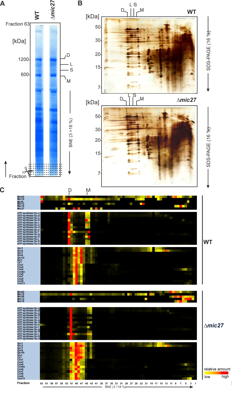
FIGURE 2: Complexome profiling of isolated mitochondria from wild type and Δmic27 cells. Mitochondria were solubilized with digitonin (ratio: digitonin to protein 2g/g) and separated with a BN-PAGE (A) or a 2D BN-PAGE/SDS-PAGE was performed (B). 1D BN-PAGE lanes (indicated in panel A) were fixed and stained with Coomassie, sectioned transversely in 63 fractions, and examined by quantitative mass spectrometry. Selected quantified proteins were represented in heat maps (C). The color scale ranges from black (not identified), to yellow (20% of the maximum), to red (maximum abundance). Monomers (M), dimers (D), and oligomers of the F1FO-ATP synthase (O) are indicated.