
Back to article: Yeast quiescence exit swiftness is influenced by cell volume and chronological age
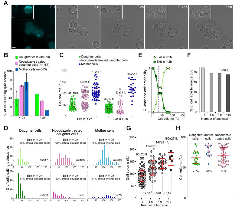
FIGURE 1: Daughter and mother cell quiescence exit efficiency. (A) Image series of 7-day-old wild type mother and daughter cells exiting quiescence on a YPD-containing microscope agarose pad. The first image shows a calcofluor white staining revealing bud scars (blue). Bar is 2 µm. (B) Percentage of 7-day-old wild type mother and daughter cells able to exit quiescence in less than 2 h (< 2 h) or between 2 and 6 h (> 2 h) on a YPD-containing microscope agarose pad at room temperature (N=3, the number of cell scored is indicated). Histograms are means and bars are SD. (C) Initial volume in function of the time needed for bud emergence. Cells are the same as in (B). Median cell volume and SD are indicated. (D) Volume distribution according to quiescence exit swiftness. Cells are the same as in (B), top graphs: emit a bud in less than 2 h; lower graphs: emit a bud within 2 to 6 h. The percentage of each sub-populations and the number of analyzed cells are indicated. (E) Probability of a daughter cell to exit quiescence in less or more than 2 h, according to its volume in quiescence. (F) Percentage and (G) volume of mother cells able to exit quiescence in less than 2 h, accordingly to their replicative age. Median cell volume and SD are indicated. (H) Cell ability to exit quiescence in less than 2 h within an identical cell volume range (50 to 90 fL). In all panels, mother cells are in blue, daughter cells in green, and nocodazole treated cells in pink.