
Back to article: Valine biosynthesis in Saccharomyces cerevisiae is regulated by the mitochondrial branched-chain amino acid aminotransferase Bat1
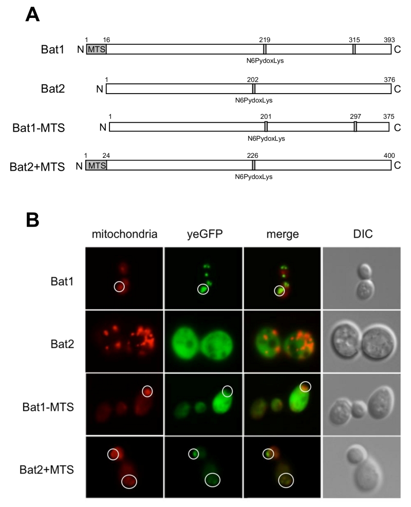
FIGURE 2: Subcellular localization of Bat1 and Bat2. (A) Schematic structure of Bat1, Bat2, Bat1-MTS, and Bat2+MTS based on UniProt [10]. (B) Subcellular localization of Bat1, Bat2, Bat1-MTS, and Bat2+MTS by fluorescent microscopy. S. cerevisiae wild-type (BY4741) cells harboring pRS416-Bat1-yeGFP/pRS415 (Bat1), pRS416-Bat1-MTS-yeGFP/pRS415 (Mat1-MTS), pRS415-Bat2-yeGFP/pRS416 (Bat2), and pRS415-Bat2+MTS-yeGFP/pRS416 (bat2+MTS) were grown in SD+His/Met medium at 30ºC for 15 h. MitoTracker signals (mitochondria), GFP signals (yeGFP), merged signals (merged), and differential interference contrast images (DIC) are shown. White circles show the mitochondria in Bat1, Bat1-MTS and Bat2+MTS.