
Back to article: Snf1 cooperates with the CWI MAPK pathway to mediate the degradation of Med13 following oxidative stress
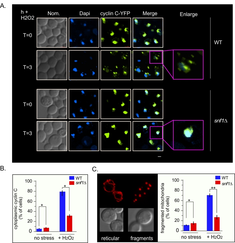
FIGURE 7: Cyclin C remains predominantly nuclear following H2O2 stress in snf1∆. (A) Fluorescence microscopy of mid-log phase wild-type and snf1∆ cells harboring a cyclin C-YFP expression plasmid (pBK38). Cells were stained with Dapi to visualize the nucleus. (B) Quantification of the results obtained in (A). At least 200 cells were counted per timepoint from 3 individual isolates. The percent of cells (mean ± SEM) within the population displaying cytoplasmic cyclin C is given. * p<0.05 difference from wild type. (C) Right panel: representative images of the two mitochondrial morphologies scored. Left panel: as in (B) except that percent of cells displaying fragmented mitochondria was scored. Representative images of the mitochondrial morphologies scored are shown in the left hand panel. The percent of cells (mean ± s.e.m.) within the population displaying fragmented mitochondria is given. * p<0.05 difference from wild type. ** p<0.01 difference from wild type. Bar = 13 µM.