
Back to article: Temporal analysis of the autophagic and apoptotic phenotypes in Leishmania parasites
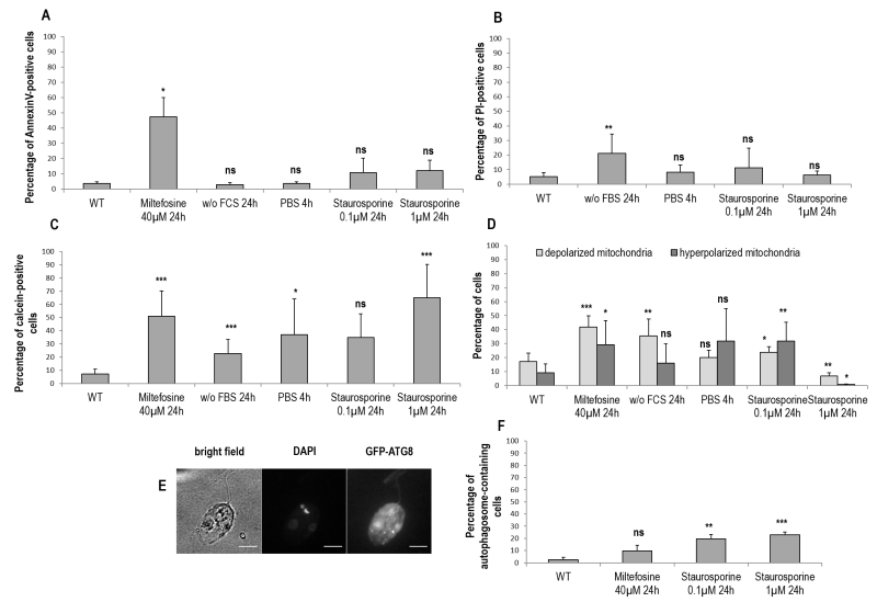
FIGURE 3: Membrane properties and autophagosome formation during Leishmania cell death and autophagy. (A) Percentage of Annexin V-positive cells in different culture conditions (≥ three independent experiments). (B) Percentage of PI-positive cells in different culture conditions (≥ three independent experiments). (C) Percentage of calcein-positive cells in different culture conditions (≥ three independent experiments). (D) Percentage of cells with a depolarized or hyperpolarized mitochondrion in different culture conditions determined by DiOC6 staining (≥ three independent experiments). (E) Microscopical observation of cells transfected with GFP-ATG8 and treated for 24 h with 0.1 µM of staurosporine (bar = 5 µm). The GFP-ATG8 puncta are indicative of autophagosomes. (F) Percentage of autophagosome-containing cells after treatment for 24 h with miltefosine and with staurosporine or not (WT). A minimum of 722 cells were counted, from three independent experiments. Unpaired t-test: ns not significant, * p < 0.05, ** p < 0.01, *** p < 0.001.