
Back to article: Temporal analysis of the autophagic and apoptotic phenotypes in Leishmania parasites
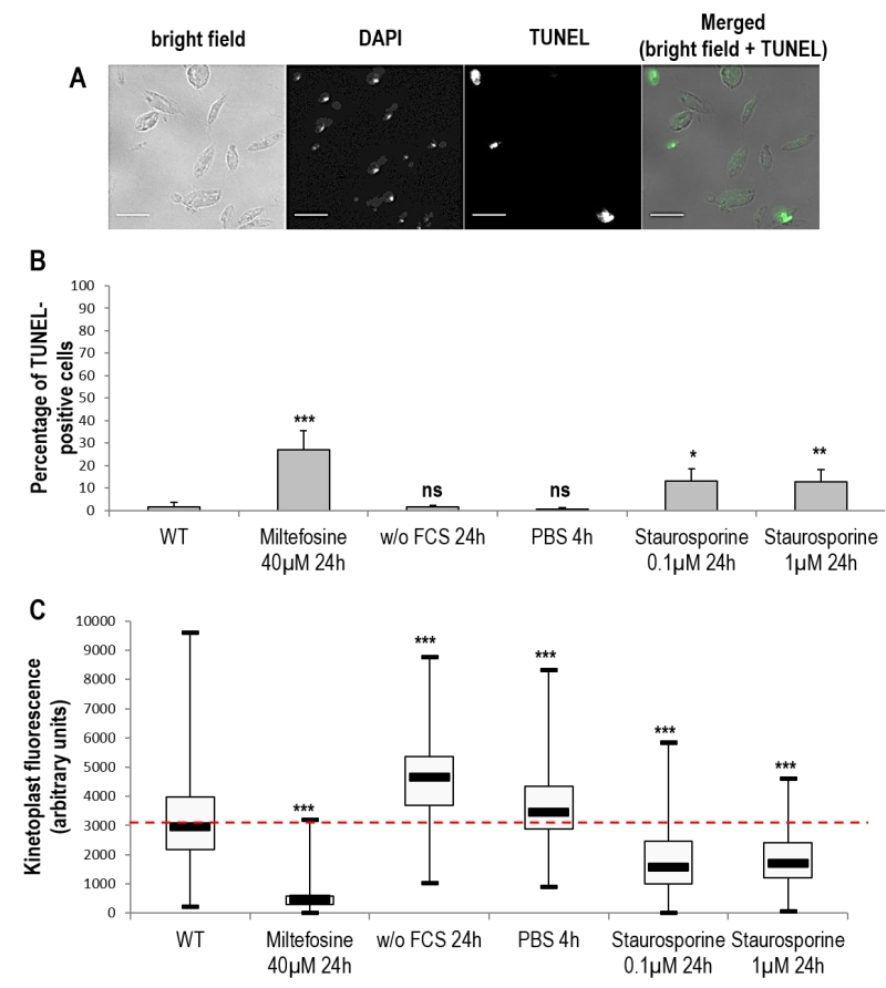
FIGURE 4: DNA degradation during Leishmania cell death and autophagy. (A) Microscopical observation of cells treated for 24 h with 0.1 µM of staurosporine and stained with DAPI and TUNEL (bar = 5 µm). TUNEL-positive nuclei indicate cells with a degraded nuclear DNA. (B) Percentage of TUNEL-positive cells in different cell culture conditions. A minimum of 1800 cells were counted from a minimum of four independent experiments. (C) Maximal kinetoplast fluorescence of cells in different culture conditions. A minimum of 200 cells were counted from minimum three independent experiments. Unpaired t-test, * p < 0.05, ** p < 0.01, *** p < 0.001.