
Back to article: Temporal analysis of the autophagic and apoptotic phenotypes in Leishmania parasites
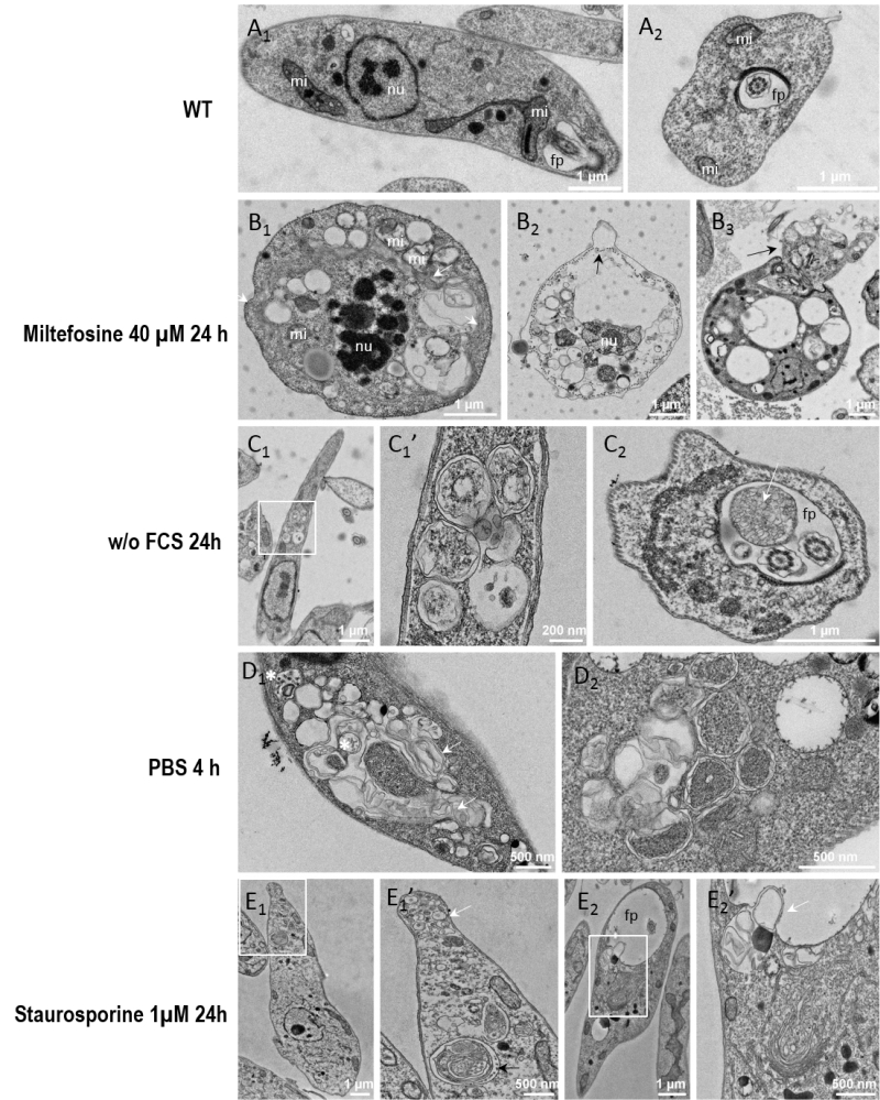
FIGURE 5: Electron micrographs of Leishmania cells during cell death and autophagy. Parasites were cultivated for 24 hours in standard conditions (A1-2), with 40 µM of miltefosine (B1-3), in a medium without FCS (C1-2), for 4 h in PBS (D1-2), or with 1 µM of staurosporine (E1-2). On images C1, E1 and E2, the white rectangle indicates a region of interest that was imaged at higher magnification shown on the next image. (A1) Longitudinal section of a normal parasite, showing the nucleus, 2 sections of the mitochondrion and the flagellar pocket containing a section of the flagellum. Note that the lumen of the flagellar pocket is electrolucent and does not show any cellular material except the flagellum. The cytoplasm does not usually contain accumulation of simple or double membrane vesicles. (A2) Transversal section through the flagellar pocket of another normal parasite. (B1) Apoptotic cell showing a shrunk nucleus with patches of highly condensed chromatin (nu) and swollen mitochondrion (mi). The plasma membrane and the subpellicular microtubule network are intact. The cytoplasm is highly vacuolated and shows accumulation of internal membrane multilayers (white arrow). (B2) Cell in late apoptosis or necrosis. Cytosolic components were essentially released, leaving degraded membranous compartments and a shrunk nucleus. Plasma membrane is making blebs (black arrow). Cortical microtubules are still visible but are not attached any longer to the plasma membrane. (B3) Apoptotic cell showing abnormal flagellar pocket partially extruding out of the cellular body (black arrow). (C1) Leishmania cells grown for 24 hours in the absence of FCS (C1) and higher magnification (C1’). An accumulation of double membrane vacuoles enclosing cytoplasmic material is observed. (C2) Cross-section of the flagellar pocket of a cell cultivated without FCS, showing a giant multivesicular body (white arrow). (D1-D2) Parasites incubated in PBS for 4 hours present accuulations of vacuoles containing membrane whorls (white arrows) and multivesicular bodies (white asterisk) (D1) and double membrane structures enclosing cytoplasm (D2). (E1: Parasites treated for 24 hours with 1µM of staurosporine contain small vesicles accumulating at the tip of the cell (white arrow). Complex multimembrane structures containing organelles and cytoplasm can also be observed in vacuoles (black arrow). (E2) The cells present a dilated flagellar pocket. A detail shown in (E2’), at higher magnification, shows a cytosolic vacuole containing membrane structures in a process of fusion or fission with the flagellar pocket. nu: nucleus; mi: mitochondrion; fp: flagellar pocket.