
Back to article: Temporal analysis of the autophagic and apoptotic phenotypes in Leishmania parasites
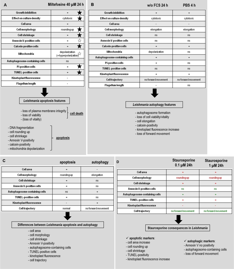
FIGURE 7: Proposed procedure to identify Leishmania apoptotic cells, autophagic cells and to clearly distinguish apoptosis and autophagy. (A) Leishmania miltefosine-induced apoptosis markers. A star indicates the apoptotic markers that have been found not only in miltefosine-induced apoptosis but also after induction of apoptosis by other molecules: a solid star when demonstrated in this article and an empty star when shown in the literature (see discussion for details). The proposed approach to demonstrate Leishmania apoptosis is also summarized. (B) Leishmania autophagy markers. The list of features observed in deprivation conditions (without FCS for 24 h and in PBS for 4 h) as well as the proposed approach to demonstrate Leishmania autophagy are summarized. (C) Features to distinguish apoptosis and autophagy in Leishmania. The characteristics that are different during Leishmania apoptosis and autophagy and so that allow distinguishing the two processes are enumerated. (D) Features induced by staurosporine in Leishmania. Staurosporine induces apoptotic (in red) as well as autophagic (in green) features in Leishmania.