
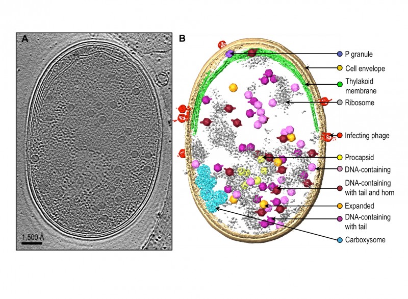
FIGURE 1: Zernike phase contrast electron cryo-tomography enables visualization of phage assembly intermediates in Syn5-infected cyanobacteria cells.
(A) Section view of a Syn5 infected cyanobacterium cell. Shown is a 54Å slab taken from the middle of the tomogram.
(B) Annotation of the tomogram. Cellular components, infecting phages and phage progeny are segmented and labelled. Intracellular phage assembly intermediates were classified into five groups based on size, shape, and density inside the capsid. Averaged maps of the five classes of phage assembly intermediates were fitted back to the original coordinates of individual intracellular particles to show the orientation of the particles.