
Back to article: D-Serine reduces the expression of the cytopathic genotoxin colibactin
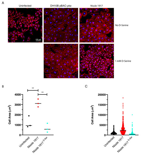
FIGURE 5: Exposure to D-Serine reduces the colibactin associated cellular senescence. HeLa cells were infected for 4 h with E. coli Nissle 1917 or DH10B hosting BAC-pks (MOI = 400). Infections were performed with and without the addition of 1 mM D-Serine to the growth media. At 8 h after infection, cells were washed and incubated for 72 h to allow for the megacell phenotype to develop. (A) HeLa cell morphology was observed by wide field fluorescence. Actin cytoskeleton was stained with Phalloidin in red and DNA was counterstained with DAPI in blue at 72 h post infection. Scale bar = 100 μm. (B) CellProfiler software was employed to measure the area of the HeLa cells shown in A using images acquired at 10X magnification. 100 cells were measured per sample. Columns represent the mean cell area measured with individual experimental observations indicated by data points for each infection condition. Measurements were acquired from images taken from three independent experiments and statistical significance was assessed by unpaired Student's t-test with, ** indicating P < 0.01. (C) Individual cell area measurements were recorded across triplicate experiments. Black lines indicate the mean.