
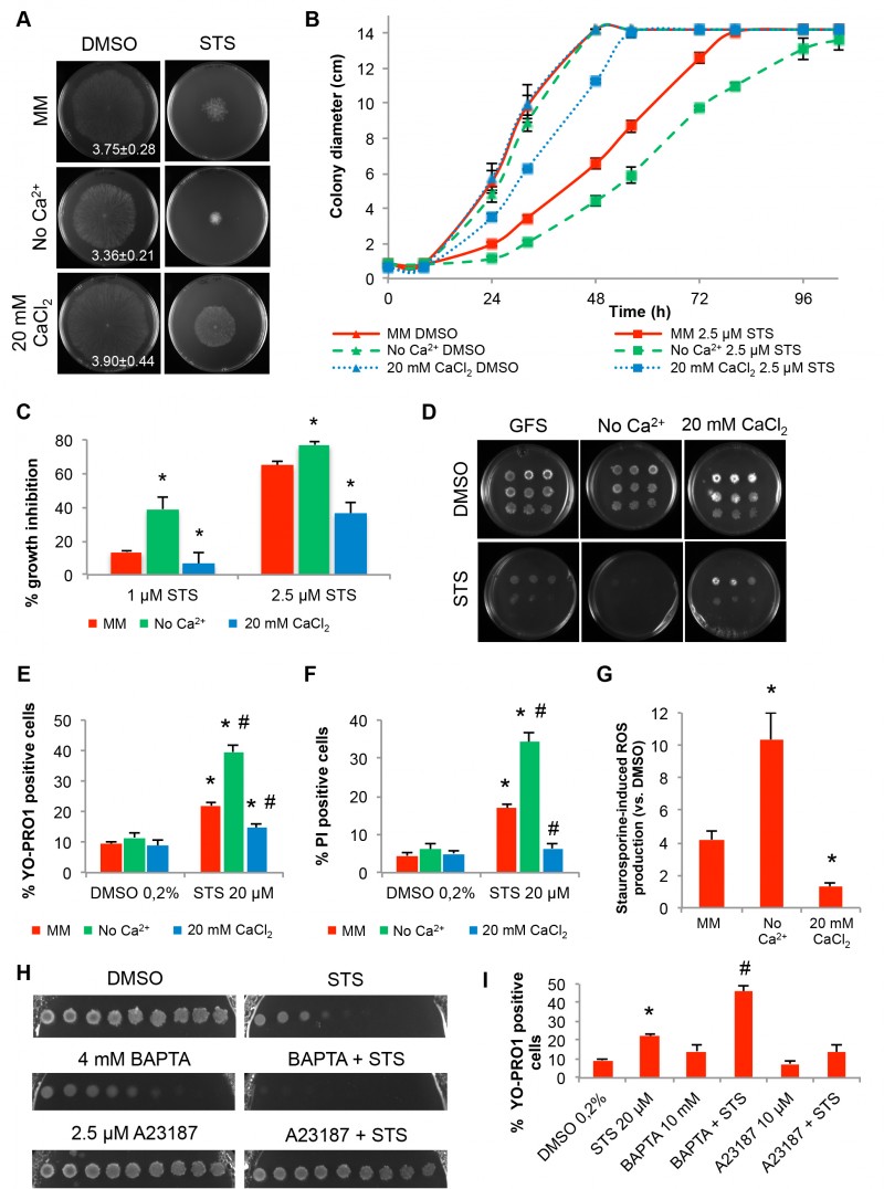
FIGURE 1: Extracellular Ca2+ modulates the N. crassa sensitivity to staurosporine.
(A-C) Conidia were inoculated on the centre of a Petri dish containing Vogel’s MM with 2.5 μM staurosporine (STS) and different concentrations of Ca2+. Growth at 32h (A), over time (B) and the percentage of growth in STS-treated cells versus control (C) are shown. Growth rates (mm/h) are indicated in the left panels in (A). *, p-value ≤ 0.05.
(D) Serial dilutions (from left to right) of conidia spotted in GFS agar medium supplemented with 5 μM STS and different concentrations of Ca2+ were incubated for 3 days.
(E-F) Cell death following treatment of the cells with 20 μM staurosporine in the indicated liquid culture media was evaluated by flow cytometry quantification of positive cells for YOPRO-1 (E) or PI (F). *, p-value ≤ 0.05 for the comparison STS versusDMSO in each media; #, p-value ≤ 0.05 for the comparison between media after the treatment with STS.
(G) The fold increase in cellular ROS accumulation following treatment with 20 μM staurosporine in the indicated liquid culture media was evaluated by staining with DHR123. *, p-value ≤ 0.05.
(H-I) Growth inhibition and cell death in 2.5 μM STS-treated cells in the presence of BAPTA or A23187 was assessed by spots in GFS medium (H) or cell growth in liquid medium followed by staining with YOPRO-1 (I) *, p-value ≤ 0.05 for the comparison STS versusDMSO; #, p-value ≤ 0.05 for the comparison between STS alone and BAPTA+STS.