
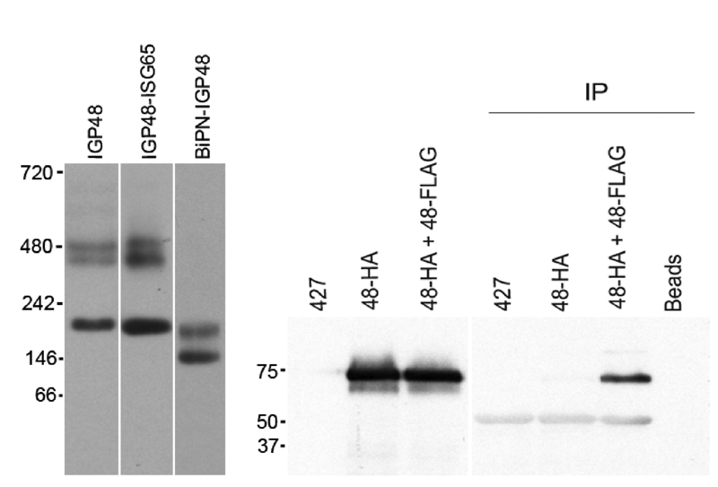
FIGURE 7: IGP48 forms a complex in vivo.
Lysates from cells expressing various IGP48 chimeras were subjected to native PAGE, followed by detection by Western blotting. Left: Replacement of the IGP48 ectodomain with the BiP ATPase domain (BiPN-IGP48) results in loss of the high molecular weight (450 – 500 kDa) complexes. Right: Cells expressing IGP48-HA or IGP48-HA (48-HA) plus IGP48-FLAG (48-FLAG) were immunoprecipitated with anti-FLAG antibody, followed by Western blotting with anti-HA antibody. Whole cell lysates are shown to the left, and wild-type (427) and single transfected cells, as well as a bead plus lysate with no antibody IP control (Beads).
For figures in higher resolution please refer to PDF version of the article.