
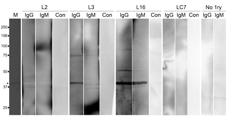
FIGURE 11: IGP48 elicits variable IgG and IgM responses in human T. b. rhodesiense infections.
Recombinant IGP48 was resolved on a 10% SDS-PAGE gel. After Western Blotting, PVDF membranes were probed with plasma from T. b. rhodesiense patients and controls.
Lane M: molecular weight markers.
Lane 1 (from left): Ponceau red staining of IGP48 (*).
Lanes 2, 3 and 4: membrane probed with plasma L2 with anti-IgG, anti-IgM and no secondary antibody control respectively.
Lanes 5, 6 and 7: membrane probed with plasma L3 with anti-IgG, anti-IgM and no secondary antibody control respectively.
Lanes 8, 9 and 10: membrane probed with plasma L16 with anti-IgG, anti-IgM and no secondary antibody control respectively.
Lanes 11, 12 and 13: membrane probed with endemic control plasma LC7 with anti-IgG, anti-IgM and no secondary antibody control respectively.
Lanes 15 and 16: anti-IgG and IgM controls with no primary antibody.
For figures in higher resolution please refer to PDF version of the article.