
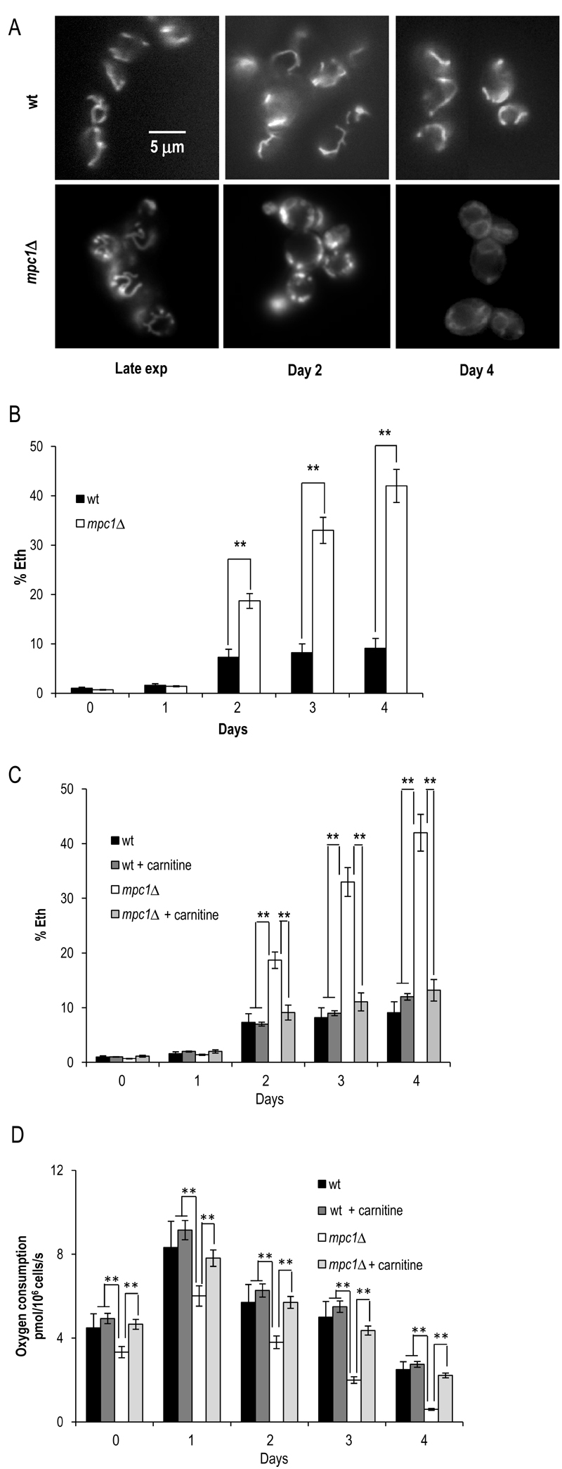
FIGURE 6: Chronologically aging mpc1∆ cells display damaged mitochondria.
(A) Representative images of wt and mpc1Δ cultures of Figure 2 stained with DiOC6 to visualize mitochondrial membranes. Morphologies of the mitochondria in late exponential phase (Late exp) are also shown.The same cultures were assessed for the presence of intracellular superoxide by conversion of non-fluorescentdihydroethidium into fluorescent ethidium (Eth). Summary graphs of the percentage of fluorescent/superoxide positive cells (% Eth) are reported (B).
(C) Summary graphs of % Eth cells and (D) cellular respiration determined in wt and mpc1Δ cultures grown in minimal medium/2% glucose supplemented with carnitine (10 mg/L).
Day 0, diauxic shift. For the determination of Eth cells, evaluation of about 1000 cells for each sample (three technical replicates) in three independent experiments was performed. SD is indicated. * P ≤ 0.05 and ** P ≤ 0.01.