
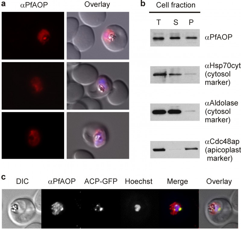
FIGURE 1: Dual localization of PfAOP.
(A) Immunofluorescence localization of PfAOP in blood stage parasites.
(B) Detection of PfAOP and marker proteins by western blotting after subcellular fractionation. The total parasite lysate (T), the supernatant (S) and the organellar pellet fraction (P) are shown from the left to the right side. PfAOP was detected at approximately 22 kDa (Fig S1). Apicoplast Cdc48, Hsp70 and aldolase were detected at approximately 130, 70 and 40 kDa, respectively.
(C) Co-localization analysis of PfAOP and a chimera of acyl-carrier protein and GFP (ACP-GFP) as an apicoplast-localized marker protein.