
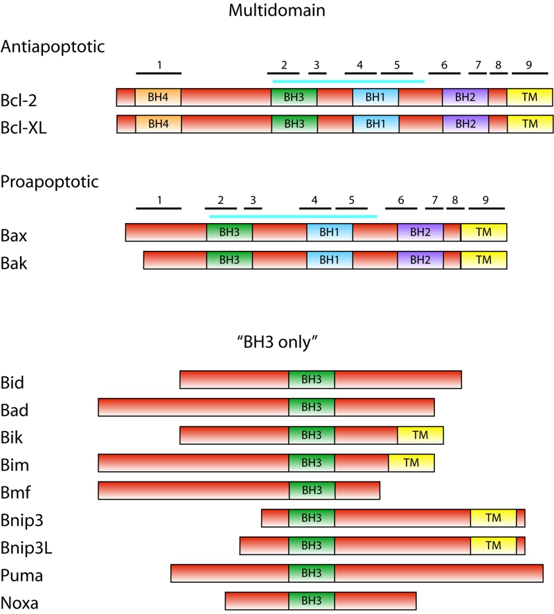
FIGURE 1: The Bcl-2 family.
Domain structure of Bcl-2 family proteins is shown schematically with sizes of proteins roughly in scale. Posi-tion of BH domains is indicated. In multidomain anti- and proapoptotic proteins, positions of α-helices are indicated with numbered black bars. Blue bar indicates the position of the hydrophobic groove. Members of the Bcl-2 family not studied in yeast and not mentioned in text are omitted.