
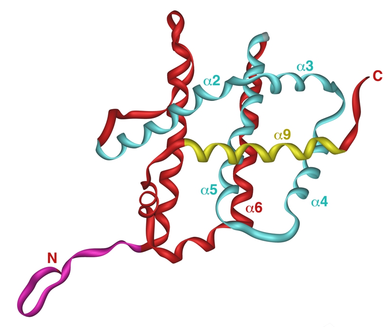
FIGURE 2: Structure of the human Bax.
NMR structure of the human Bax is shown with following structural features emphasized: N-terminal domain in purple, hydrophobic groove in blue and α-helix 9 in yellow. Picture was generated by iMol 0.40 software using structural data from [57].
57. Suzuki M, Youle RJ and Tjandra N (2000). Structure of Bax: coregulation of dimer formation and intra-cellular localization. Cell. 103(4): 645-654. http://dx.doi.org/10.1016/s0092-8674(00)00167-7