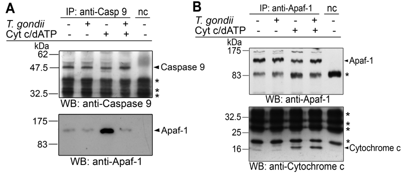
FIGURE 4: Interference of T. gondii with holo-apoptosome assembly as revealed by co-immunoprecipitation analyses.
(A, B) Cell-free cytosolic extracts from Jurkat cells were incubated or not with T. gondii (108/ml). After 1 hour, apoptosome formation was triggered by addition of cytochrome c and dATP as indicated. Caspase 9 (A) or Apaf-1 (B) was immunoprecipitated using specific antibodies and protein A-sepharose in the presence of a caspase 3-inhibitor. A negative control precipitation without cell lysate was run in parallel (nc). Precipitates were resolved by SDS-PAGE and were analyzed by immunoblotting using specific antibodies as indicated. Bound antibodies were visualized by enhanced chemiluminescence after incubation with appropriate peroxidase-conjugated secondary antibodies. Unspecific binding of secondary antibodies is indicated by asterisks. The experiment was repeated twice with similar results.

