
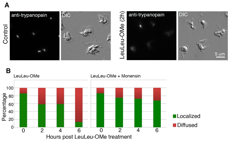
FIGURE 3: LeuLeu-OMe-induced lysosome destabilization revealed by anti-trypanopain staining.
(A) Control and LeuLeu-OMe-treated cells were fixed and stained with anti-trypanopain. The staining was localized to one or two puncta in control cells, but became diffused in LeuLeu-OMe-treated cells. Control and drug-treated cells were processed for immunostaining using same conditions. Note that LeuLeu-OMe-treated cells were exposed longer to capture the weaker anti-trypanopain staining in the cytosol.
(B) Cells with localized (green) or diffused (red) anti-trypanopain staining were quantitated over the course of LeuLeu-OMe treatment (left), and in cells pre-treated with monensin (right). At least 200 cells were counted for each time point.