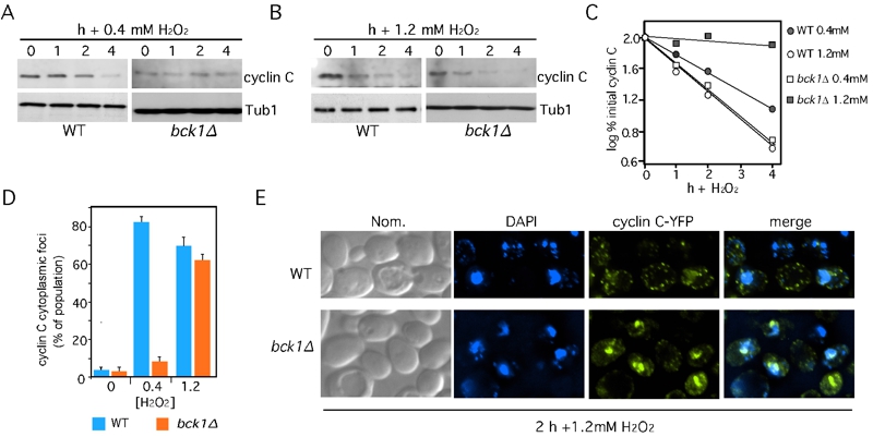
FIGURE 2: Bck1 is not required for cyclin C nuclear to cytoplasmic translocation and degradation following high (1.2 mM) H2O2 stress.
Wild type (RSY10) and bck1∆ (RSY1050) cultures expressing myc-cyclin C (pLR337) were grown to mid-log phase (0 h) then treated with 0.4 mM (A) or 1.2 mM (B) H2O2 for the indicated times. Cyclin C levels were determined by Western blot analysis of immunoprecipitates. Tub1 levels were used as a loading control.
(C) Quantification of the results obtained in (A) and (B).
(D) The percent of cells (mean ± s.e.m.) within the population displaying at least 3 cytoplasmic cyclin C foci is given before and following H2O2 (0.4 and 1.2 mM) treatment for 2 h. At least 200 cells were counted per time point from 3 individual isolates.
(E) Representative images (collapsed de-convolved 2 µm slices) of WT and bck1∆ cells harboring cyclin C-YFP and after 2 h 1.2 mM H2O2 stress.

