
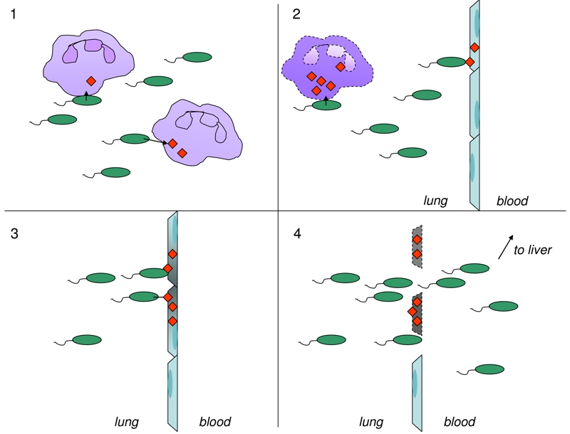
FIGURE 1: Model for ExoS-mediated bacterial dissemination during acute pneumonia.
1. Early during infection, neutrophils (purple) are injected with ExoS (red), preventing phagocytosis and clearance of P. aeruginosa bacteria (green).
2. Later during infection, bacteria encounter the pulmonary-vascular barrier and inject type I pneumocytes (blue) with ExoS.
3. ExoS injection occurs at discrete regions termed FOCI (gray), which expand as infection progresses and type I pneumocytes are killed.
4. Pulmonary-vascular leakage allows bacteria to disseminate into the bloodstream and to the liver.