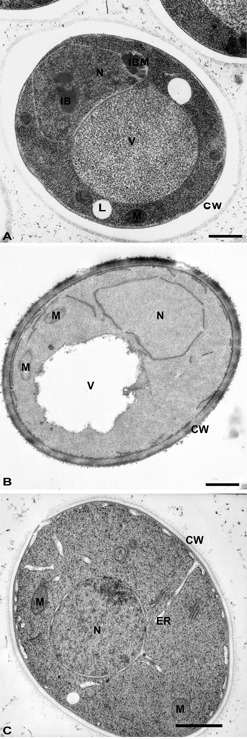
FIGURE 2: Morphology of yeast cells embedded in epoxy resins.
(A) Cells were cryofixed in liquid propane, freeze-substituted in acetone containing 4% OsO4 and embedded in Epon. CW, cell wall; N, Nucleus; IB, Inclusion body; IBM, Inclusion body with membrane; L, lipid droplets; V, Vacuole. Scale bar, 0.5 µm. This image was originally published in [157] © Springer.
(B) Yeast was fixed with 1.5% KMnO4, dehydrated with acetone and embedded in Spurr’s resin. CW, cell wall; M, mitochondria; N, Nucleus; V, vacuole. Scale bar, 0.5 µm. This image was originally published in [158] © the American Society for Biochemistry and Molecular Biology.
(C) Cells were high-pressure frozen, freeze-substituted in acetone, and embedded in a mixture of Epon-Spurr’s resin. CW, cell wall; ER, endoplasmic reticulum; M, mitochondria; N, nucleus. Scale Bar, 1.0 µm. This image was originally published in [26] © Elsevier Limited.
26. McDonald K. (2007). Cryopreparation methods for electron microscopy of selected model systems. Methods Cell Biol. 7923-56. http://dx.doi.org/S0091-679X(06)79002-1
157. Binder M, Hartig AT. (1996). Immunogold labeling of yeast cells: an efficient tool for the study of protein targeting and morphological alterations due to overexpression and inactivation of genes. Histochem.Cell Biol. 106(1): 115-130. http://dx.doi.org/10.1007/BF02473206
158. Cabrera M, Arlt H, Epp N, Lachmann J, Griffith J, Perz A, Reggiori F, Ungermann C. (2013). Functional separation of endosomal fusion factors and the class C core vacuole/endosome tethering (CORVET) complex in endosome biogenesis. J.Biol.Chem. 288(7): 5166-5175. http://dx.doi.org/10.1074/jbc.M112.431536

