
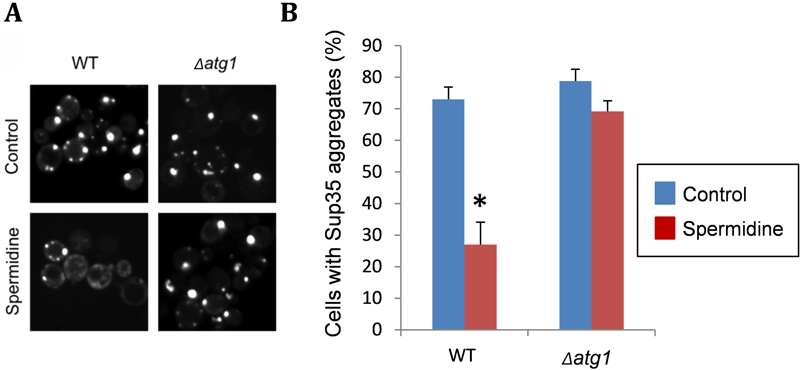
FIGURE 2: Spermidine treatment decreases the number of cells with visible Sup35 fluorescent aggregates.
(A) Representative fluorescence micrographs are shown for [PIN+][PSI+] versions of the wild-type yeast strain 74D-694 (MATa ade1-14 ura3-52 leu2-3,112 trp1-289 his3-200) and an isogenic atg1 mutant containing the Sup35NM-GFP plasmid. Strains were grown in minimal media in the presence or absence of 4 mM spermidine for 48 hours to induce autophagy. Sup35NM-GFP was induced with copper for one hour. Following copper induction, fluorescent foci can be detected due to the coalescence of newly made Sup35NM-GFP with pre-existing Sup35 aggregates.
(B) The percentage of cells containing visible puncta is shown for each strain from an average of 300 cells counted. Data shown are the means of three independent biological repeat experiments ± SD. The number of visible aggregates in the wild-type strain treated with spermidine is significantly different to the number of aggregates detected in the same strain in the absence of spermidine (*p = <0.001).