
Back to article: A multigene family encoding surface glycoproteins in Trypanosoma congolense
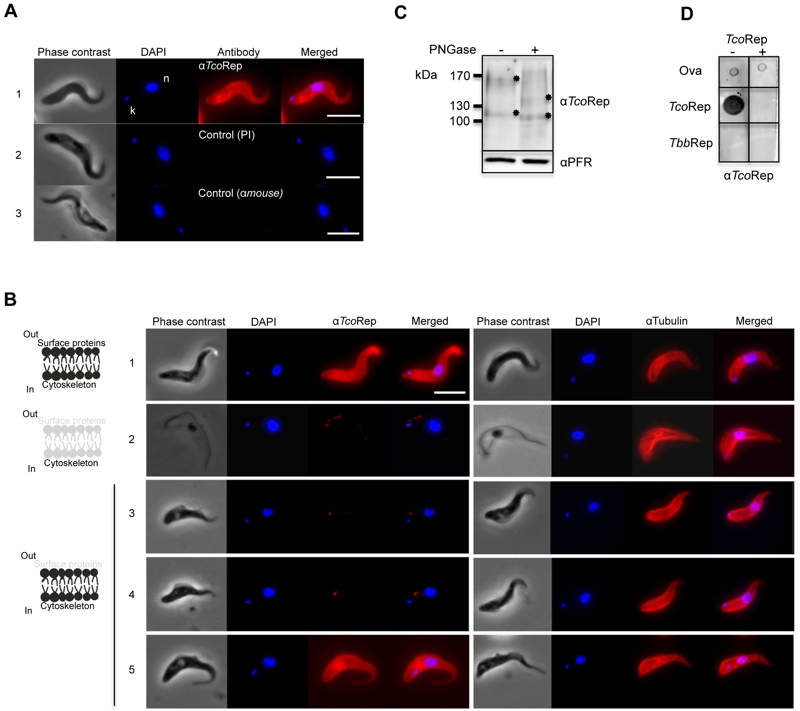
FIGURE 3: Expression and cellular localization of TcoClec in BSF of T. congolense.
(A) Immunofluorescence analysis of T. congolense BSF. Cells were stained with mouse anti-TcoRep (1); control pre-immune serum (2); or secondary antibody alone (3). DAPI was used to stain both nucleus (n) and kinetoplast (k). Phase contrast images are shown on the left.
(B) Immunofluorescence analysis. Images are typical examples showing phase contrast, DAPI staining of the nucleus and the kinetoplast, and staining with anti-TcoRep or anti-tubulin. Whole-cell T. congolense BSF (1); cytoskeleton-extracted cells (treated with NP40) (2); and tryp-sin-treated cells (« shaved » parasites) with IFA performed immediately (3), 5 h (4) and 14 h (5) after the treatment. A schematic illustration is shown on the left to explain the effects of the treatments on Tco BSF. Bar, 5 µm.
(C) Western blot analysis. Tco BSF cell lysates treated (+) or not (-) with PNGase F. Mouse anti-TcoRep and rabbit anti-PFR (control) were used. Asterisks show band shifts.
(D) Peptide competition assay (PCA). Dot blot analysis developed with anti-TcoRep antibodies. Anti-TcoRep antibodies were incubated (+) or not (-) with the TcoRep peptide. Ovalbu-min (Ova), TcoRep peptide and TbbRep peptide were spotted on the filters.