Table of contents
Volume 5, Issue 6, pp. 262 - 299, June 2018
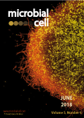
Cover: Streptomyces lividans 66 producing poly-N-acetylglucosamine (PNAG). The image shows a mycelium from an 18 h old liquid-grown culture of the matAB double mutant genetically complemented by introduction of a plasmid expressing matAB. PNAG was stained with monoclonal antibody mAb F598 (yellow). To visualize the hyphae, the DNA was stained with PI (red). Image by Dino van Dissel and Gilles P. van Wezel (Leiden University, The Netherlands); image modified by MIC. The cover is published under the Creative Commons Attribution (CC BY) license.
Enlarge issue cover
The CRISPR conundrum: evolve and maybe die, or survive and risk stagnation
Jesús García-Martínez, Rafael D. Maldonado, Noemí M. Guzmán and Francisco J. M. Mojica
Reviews |
page 262-268 | 10.15698/mic2018.06.634 | Full text | PDF |
Abstract
CRISPR-Cas represents a prokaryotic defense mechanism against invading genetic elements. Although there is a diversity of CRISPR-Cas systems, they all share similar, essential traits. In general, a CRISPR-Cas system consists of one or more groups of DNA repeats named CRISPR (Clustered Regularly Interspaced Short Palindromic Repeats), regularly separated by unique sequences referred to as spacers, and a set of functionally associated cas (CRISPR associated) genes typically located next to one of the repeat arrays. The origin of spacers is in many cases unknown but, when ascertained, they usually match foreign genetic molecules. The proteins encoded by some of the cas genes are in charge of the incorporation of new spacers upon entry of a genetic element. Other Cas proteins participate in generating CRISPR-spacer RNAs and perform the task of destroying nucleic acid molecules carrying sequences similar to the spacer. In this way, CRISPR-Cas provides protection against genetic intruders that could substantially affect the cell viability, thus acting as an adaptive immune system. However, this defensive action also hampers the acquisition of potentially beneficial, horizontally transferred genes, undermining evolution. Here we cover how the model bacterium Escherichia coli deals with CRISPR-Cas to tackle this major dilemma, evolution versus survival.
Production of poly-β-1,6-N-acetylglucosamine by MatAB is required for hyphal aggregation and hydrophilic surface adhesion by Streptomyces
Dino van Dissel, Joost Willemse, Boris Zacchetti, Dennis Claessen, Gerald B. Pier, Gilles P. van Wezel
Research Articles |
page 269-279 | 10.15698/mic2018.06.635 | Full text | PDF |
Abstract
Streptomycetes are multicellular filamentous microorganisms, and major producers of industrial enzymes and bioactive compounds such as antibiotics and anticancer drugs. The mycelial lifestyle plays an important role in the productivity during industrial fermentations. The hyphae of liquid-grown streptomycetes can self-aggregate into pellets, which hampers their industrial exploitation. Here we show that the Mat complex, which is required for pellet formation, catalyzes the synthesis of extracellular poly-β-1,6-N-acetylglucosamine (PNAG) in the model organisms Streptomyces coelicolor and Streptomyces lividans. Extracellular accumulation of PNAG allows Streptomyces to attach to hydrophilic surfaces, while attachment to hydrophobic surfaces requires a cellulase-degradable extracellular polymer (EPS) produced by CslA. Over-expression of matAB was sufficient to restore pellet formation to cslA null mutants of S. lividans. The two EPS systems together increase the robustness of mycelial pellets. These new insights allow better control of liquid-culture morphology of streptomycetes, which may be harnessed to improve growth and industrial exploitation of these highly versatile natural product and enzyme producers.
Evolution of substrate specificity in the Nucleobase-Ascorbate Transporter (NAT) protein family
Anezia Kourkoulou, Alexandros A. Pittis and George Diallinas
Research Reports |
page 280-292 | 10.15698/mic2018.06.636 | Full text | PDF |
Abstract
L-ascorbic acid (vitamin C) is an essential metabolite in animals and plants due to its role as an enzyme co-factor and antioxidant activity. In most eukaryotic organisms, L-ascorbate is biosynthesized enzymatically, but in several major groups, including the primate suborder Haplorhini, this ability is lost due to gene truncations in the gene coding for L-gulonolactone oxidase. Specific ascorbate transporters (SVCTs) have been characterized only in mammals and shown to be essential for life. These belong to an extensively studied transporter family, called Nucleobase-Ascorbate Transporters (NAT). The prototypic member of this family, and one of the most extensively studied eukaryotic transporters, is UapA, a uric acid-xanthine/H+ symporter in the fungus Aspergillus nidulans. Here, we investigate molecular aspects of NAT substrate specificity and address the evolution of ascorbate transporters apparently from ancestral nucleobase transporters. We present a phylogenetic analysis, identifying a distinct NAT clade that includes all known L-ascorbate transporters. This clade includes homologues only from vertebrates, and has no members in non-vertebrate or microbial eukaryotes, plants or prokaryotes. Additionally, we identify within the substrate-binding site of NATs a differentially conserved motif, which we propose is critical for nucleobase versus ascorbate recognition. This conclusion is supported by the amino acid composition of this motif in distinct phylogenetic clades and mutational analysis in the UapA transporter. Together with evidence obtained herein that UapA can recognize with extremely low affinity L-ascorbate, our results support that ascorbate-specific NATs evolved by optimization of a sub-function of ancestral nucleobase transporters.
Valine biosynthesis in Saccharomyces cerevisiae is regulated by the mitochondrial branched-chain amino acid aminotransferase Bat1
Natthaporn Takpho, Daisuke Watanabe and Hiroshi Takagi
Research Reports |
page 293-299 | 10.15698/mic2018.06.637 | Full text | PDF |
Abstract
In the yeast Saccharomyces cerevisiae, the branched-chain amino acid aminotransferases (BCATs) Bat1 and Bat2 catalyze the conversion of α-ketoisovalerate, α-keto-β-methylvalerate, and α-ketoisokaproate and into valine, isoleucine, and leucine, respectively, as the final step of branched-chain amino acid biosynthesis. Bat1 and Bat2 are homologous proteins that share 77% identity, but Bat1 localizes in the mitochondria and Bat2 in the cytosol. Based on our preliminary finding that only disruption of the BAT1 gene led to slow-growth phenotype, we hypothesized that Bat1 and Bat2 play distinct roles in valine biosynthesis and the regulation of cell growth. In this study, we found that intracellular valine content was dramatically decreased in Δbat1 cells, whereas Δbat2 cells exhibited no changes in the valine level. To further examine the distinct roles of Bat1 and Bat2, we constructed two artificial genes encoding the mitochondrial-targeting signal (MTS)-deleted Bat1 (Bat1-MTS) and the MTS of Bat1-fused Bat2 (Bat2+MTS). Interestingly, Bat2+MTS was relocalized into the mitochondria, because Bat2 localization was changed to the mitochondria by addition of MTS, and could partially restore the valine content and growth in Δbat1Δbat2 cells. These results suggest that the mitochondria are the major site of valine biosynthesis, and mitochondrial BCAT is important for valine biosynthesis in S. cerevisiae.


