Back to article: A humanized yeast-based toolkit for monitoring phosphatidylinositol 3-kinase activity at both single cell and population levels
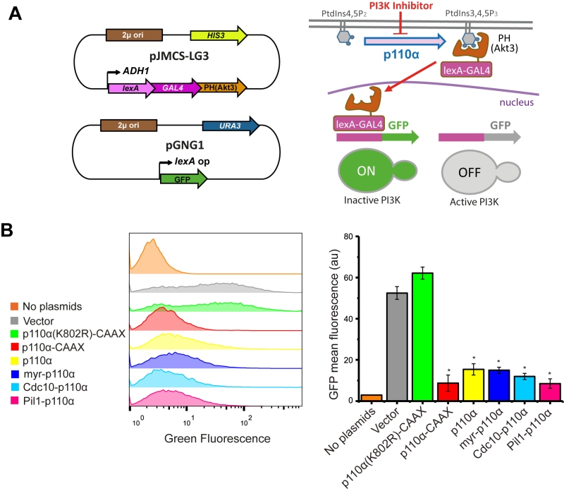
FIGURE 4: The FLUorescence by PI3K Inhibition (FLUPI) assay for the detection of heterologous PI3K activity by flow cytometry. (A) Sketches of plasmid pJMCS-LG3 bearing the transcriptional activator construct and pGNG1 (Mobitec) (left) and rationale of the system (right): the LexA-Gal4-PH(Akt3) reporter is retained at the PM when the PI3K product PtdIns3,4,5P3 is present. Its release due to low PI3K activity or its inhibition should proportionally lead to nuclear translocation of the chimeric transcription factor and GFP expression. (B) On the left, stacked histograms (n = 10,000) showing green fluorescence in abscissae of untransformed YPH499 strain (No plasmids) and triple co-transformant clones bearing pJMCS-LG3, pGNG1 and either empty YCpLG (Vector) or expressing one of the PI3K versions: p110α(K802R)-CAAX, p110α-CAAX, p110α, myr-p110α, Cdc10-p110α or Pil1-p110α. On the right, graph showing each population´s mean GFP fluorescence (au) corresponding to the average of three biological replicates (n = 30,000). The asterisks (*) indicate a Bonferroni p-value < 0.001 vs. Vector; error bars represent SD.

